Nickel »
PDB 3qsi-3tsn »
3rdo »
Nickel in PDB 3rdo: Crystal Structure of R7-2 Streptavidin Complexed with Biotin
Protein crystallography data
The structure of Crystal Structure of R7-2 Streptavidin Complexed with Biotin, PDB code: 3rdo
was solved by
V.N.Malashkevich,
M.Magalhaes,
C.M.Czecster,
R.Guan,
M.Levy,
S.C.Almo,
with X-Ray Crystallography technique. A brief refinement statistics is given in the table below:
| Resolution Low / High (Å) | 19.26 / 1.40 |
| Space group | I 41 2 2 |
| Cell size a, b, c (Å), α, β, γ (°) | 57.360, 57.360, 184.067, 90.00, 90.00, 90.00 |
| R / Rfree (%) | 15.1 / 17.1 |
Nickel Binding Sites:
The binding sites of Nickel atom in the Crystal Structure of R7-2 Streptavidin Complexed with Biotin
(pdb code 3rdo). This binding sites where shown within
5.0 Angstroms radius around Nickel atom.
In total only one binding site of Nickel was determined in the Crystal Structure of R7-2 Streptavidin Complexed with Biotin, PDB code: 3rdo:
In total only one binding site of Nickel was determined in the Crystal Structure of R7-2 Streptavidin Complexed with Biotin, PDB code: 3rdo:
Nickel binding site 1 out of 1 in 3rdo
Go back to
Nickel binding site 1 out
of 1 in the Crystal Structure of R7-2 Streptavidin Complexed with Biotin
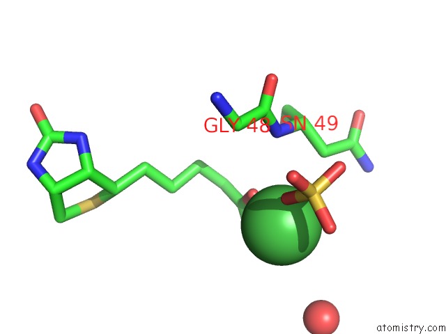
Mono view
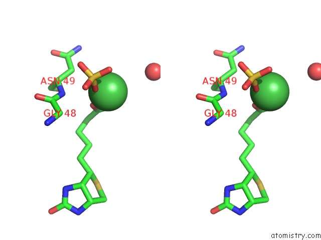
Stereo pair view
Mono view
Stereo pair view
A full contact list of Nickel with other atoms in the Ni binding
site number 1 of Crystal Structure of R7-2 Streptavidin Complexed with Biotin within 5.0Å range:
|
Reference:
M.L.Magalhaes,
C.M.Czekster,
R.Guan,
V.N.Malashkevich,
S.C.Almo,
M.Levy.
Evolved Streptavidin Mutants Reveal Key Role of Loop Residue in High-Affinity Binding. Protein Sci. V. 20 1145 2011.
ISSN: ISSN 0961-8368
PubMed: 21520321
DOI: 10.1002/PRO.642
Page generated: Wed Oct 9 17:45:44 2024
ISSN: ISSN 0961-8368
PubMed: 21520321
DOI: 10.1002/PRO.642
Last articles
Fe in 2YXOFe in 2YRS
Fe in 2YXC
Fe in 2YNM
Fe in 2YVJ
Fe in 2YP1
Fe in 2YU2
Fe in 2YU1
Fe in 2YQB
Fe in 2YOO