Nickel »
PDB 4ofo-4rro »
4qdw »
Nickel in PDB 4qdw: Joint X-Ray and Neutron Structure of Streptomyces Rubiginosus D-Xylose Isomerase in Complex with Two NI2+ Ions and Linear L-Arabinose
Enzymatic activity of Joint X-Ray and Neutron Structure of Streptomyces Rubiginosus D-Xylose Isomerase in Complex with Two NI2+ Ions and Linear L-Arabinose
All present enzymatic activity of Joint X-Ray and Neutron Structure of Streptomyces Rubiginosus D-Xylose Isomerase in Complex with Two NI2+ Ions and Linear L-Arabinose:
5.3.1.5;
5.3.1.5;
Protein crystallography data
The structure of Joint X-Ray and Neutron Structure of Streptomyces Rubiginosus D-Xylose Isomerase in Complex with Two NI2+ Ions and Linear L-Arabinose, PDB code: 4qdw
was solved by
A.Y.Kovalevsky,
P.Langan,
with X-Ray Crystallography technique. A brief refinement statistics is given in the table below:
| Resolution Low / High (Å) | 20.00 / 1.80 |
| Space group | I 2 2 2 |
| Cell size a, b, c (Å), α, β, γ (°) | 94.190, 99.670, 102.940, 90.00, 90.00, 90.00 |
| R / Rfree (%) | 16.6 / 17.9 |
Nickel Binding Sites:
The binding sites of Nickel atom in the Joint X-Ray and Neutron Structure of Streptomyces Rubiginosus D-Xylose Isomerase in Complex with Two NI2+ Ions and Linear L-Arabinose
(pdb code 4qdw). This binding sites where shown within
5.0 Angstroms radius around Nickel atom.
In total 3 binding sites of Nickel where determined in the Joint X-Ray and Neutron Structure of Streptomyces Rubiginosus D-Xylose Isomerase in Complex with Two NI2+ Ions and Linear L-Arabinose, PDB code: 4qdw:
Jump to Nickel binding site number: 1; 2; 3;
In total 3 binding sites of Nickel where determined in the Joint X-Ray and Neutron Structure of Streptomyces Rubiginosus D-Xylose Isomerase in Complex with Two NI2+ Ions and Linear L-Arabinose, PDB code: 4qdw:
Jump to Nickel binding site number: 1; 2; 3;
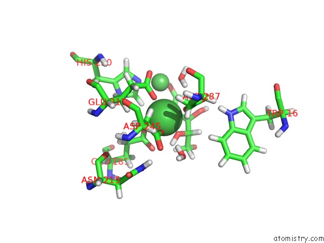
Nickel binding site 1 out of 3 in 4qdw
Go back to
Nickel binding site 1 out
of 3 in the Joint X-Ray and Neutron Structure of Streptomyces Rubiginosus D-Xylose Isomerase in Complex with Two NI2+ Ions and Linear L-Arabinose
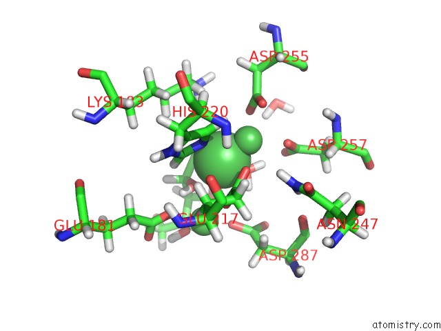
Mono view
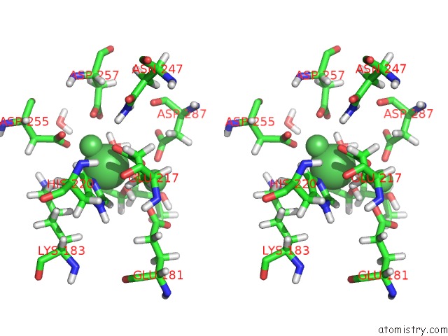
Stereo pair view
Mono view
Stereo pair view
A full contact list of Nickel with other atoms in the Ni binding
site number 1 of Joint X-Ray and Neutron Structure of Streptomyces Rubiginosus D-Xylose Isomerase in Complex with Two NI2+ Ions and Linear L-Arabinose within 5.0Å range:
|
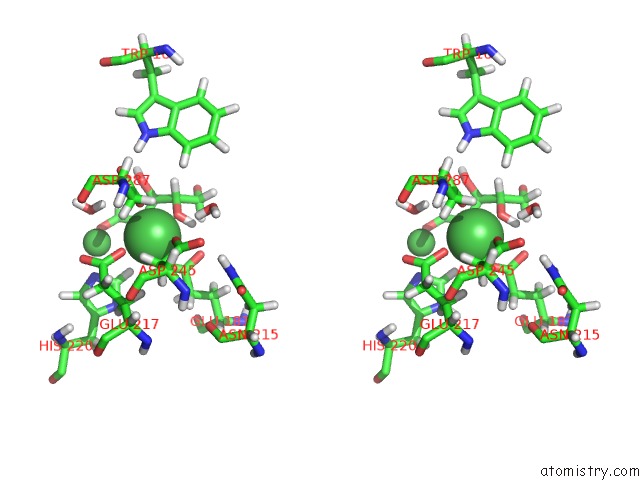
Nickel binding site 2 out of 3 in 4qdw
Go back to
Nickel binding site 2 out
of 3 in the Joint X-Ray and Neutron Structure of Streptomyces Rubiginosus D-Xylose Isomerase in Complex with Two NI2+ Ions and Linear L-Arabinose
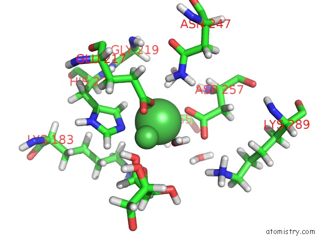
Mono view
Stereo pair view
Mono view
Stereo pair view
A full contact list of Nickel with other atoms in the Ni binding
site number 2 of Joint X-Ray and Neutron Structure of Streptomyces Rubiginosus D-Xylose Isomerase in Complex with Two NI2+ Ions and Linear L-Arabinose within 5.0Å range:
|
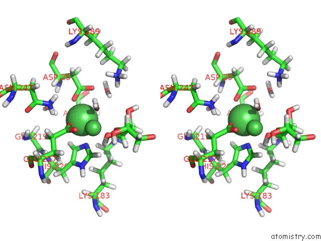
Nickel binding site 3 out of 3 in 4qdw
Go back to
Nickel binding site 3 out
of 3 in the Joint X-Ray and Neutron Structure of Streptomyces Rubiginosus D-Xylose Isomerase in Complex with Two NI2+ Ions and Linear L-Arabinose
Mono view
Stereo pair view
Mono view
Stereo pair view
A full contact list of Nickel with other atoms in the Ni binding
site number 3 of Joint X-Ray and Neutron Structure of Streptomyces Rubiginosus D-Xylose Isomerase in Complex with Two NI2+ Ions and Linear L-Arabinose within 5.0Å range:
|
Reference:
P.Langan,
A.K.Sangha,
T.Wymore,
J.M.Parks,
Z.K.Yang,
B.L.Hanson,
Z.Fisher,
S.A.Mason,
M.P.Blakeley,
V.T.Forsyth,
J.P.Glusker,
H.L.Carrell,
J.C.Smith,
D.A.Keen,
D.E.Graham,
A.Kovalevsky.
L-Arabinose Binding, Isomerization, and Epimerization By D-Xylose Isomerase: X-Ray/Neutron Crystallographic and Molecular Simulation Study. Structure V. 22 1287 2014.
ISSN: ISSN 0969-2126
PubMed: 25132082
DOI: 10.1016/J.STR.2014.07.002
Page generated: Wed Oct 9 18:39:38 2024
ISSN: ISSN 0969-2126
PubMed: 25132082
DOI: 10.1016/J.STR.2014.07.002
Last articles
Fe in 2YXOFe in 2YRS
Fe in 2YXC
Fe in 2YNM
Fe in 2YVJ
Fe in 2YP1
Fe in 2YU2
Fe in 2YU1
Fe in 2YQB
Fe in 2YOO