Nickel »
PDB 5tvr-5xgz »
5vb0 »
Nickel in PDB 5vb0: Crystal Structure of Fosfomycin Resistance Protein FOSA3
Protein crystallography data
The structure of Crystal Structure of Fosfomycin Resistance Protein FOSA3, PDB code: 5vb0
was solved by
E.Klontz,
S.Guenther,
Z.Silverstein,
E.Sundberg,
with X-Ray Crystallography technique. A brief refinement statistics is given in the table below:
| Resolution Low / High (Å) | 29.75 / 2.69 |
| Space group | P 41 2 2 |
| Cell size a, b, c (Å), α, β, γ (°) | 87.608, 87.608, 357.038, 90.00, 90.00, 90.00 |
| R / Rfree (%) | 20.5 / 24.9 |
Other elements in 5vb0:
The structure of Crystal Structure of Fosfomycin Resistance Protein FOSA3 also contains other interesting chemical elements:
| Manganese | (Mn) | 8 atoms |
Nickel Binding Sites:
The binding sites of Nickel atom in the Crystal Structure of Fosfomycin Resistance Protein FOSA3
(pdb code 5vb0). This binding sites where shown within
5.0 Angstroms radius around Nickel atom.
In total 6 binding sites of Nickel where determined in the Crystal Structure of Fosfomycin Resistance Protein FOSA3, PDB code: 5vb0:
Jump to Nickel binding site number: 1; 2; 3; 4; 5; 6;
In total 6 binding sites of Nickel where determined in the Crystal Structure of Fosfomycin Resistance Protein FOSA3, PDB code: 5vb0:
Jump to Nickel binding site number: 1; 2; 3; 4; 5; 6;
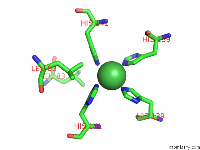
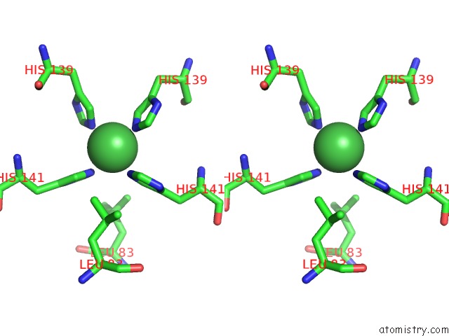
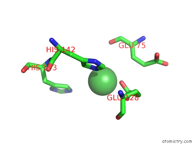
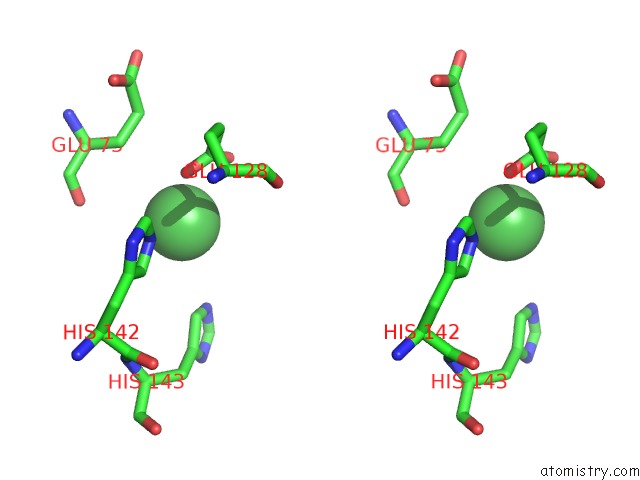
Nickel binding site 1 out of 6 in 5vb0
Go back to
Nickel binding site 1 out
of 6 in the Crystal Structure of Fosfomycin Resistance Protein FOSA3
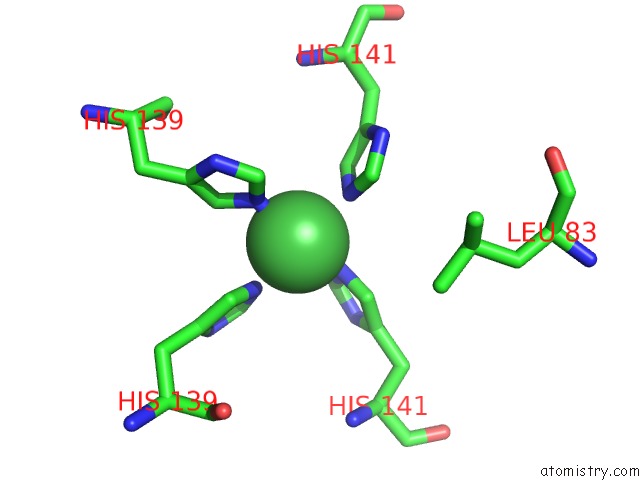
Mono view
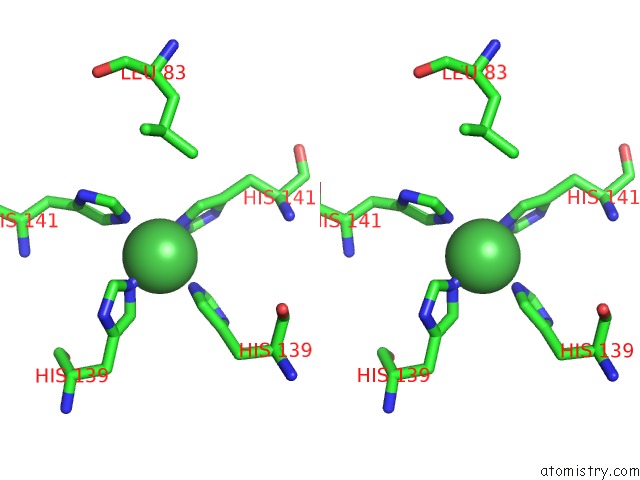
Stereo pair view
Mono view
Stereo pair view
A full contact list of Nickel with other atoms in the Ni binding
site number 1 of Crystal Structure of Fosfomycin Resistance Protein FOSA3 within 5.0Å range:
|
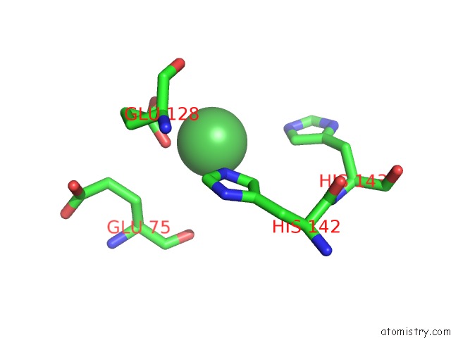
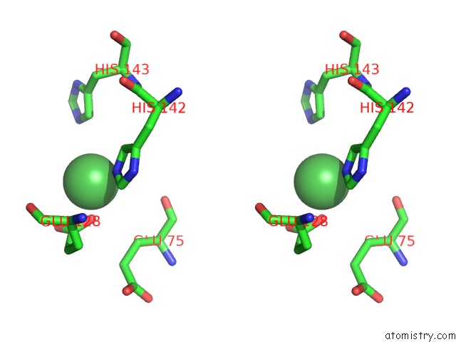
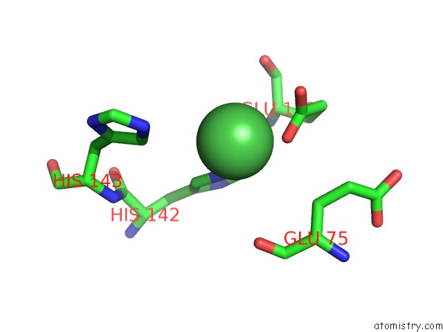
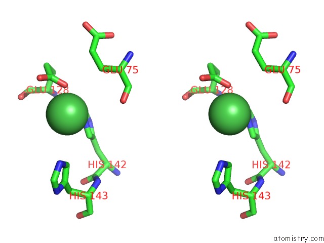
Nickel binding site 2 out of 6 in 5vb0
Go back to
Nickel binding site 2 out
of 6 in the Crystal Structure of Fosfomycin Resistance Protein FOSA3
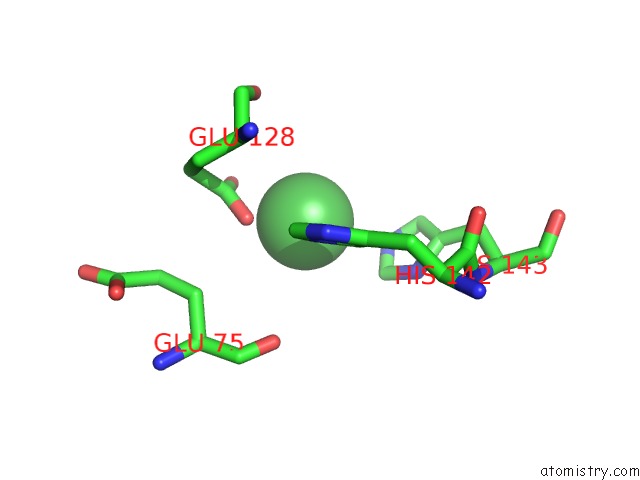
Mono view
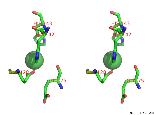
Stereo pair view
Mono view
Stereo pair view
A full contact list of Nickel with other atoms in the Ni binding
site number 2 of Crystal Structure of Fosfomycin Resistance Protein FOSA3 within 5.0Å range:
|
Nickel binding site 3 out of 6 in 5vb0
Go back to
Nickel binding site 3 out
of 6 in the Crystal Structure of Fosfomycin Resistance Protein FOSA3
Mono view
Stereo pair view
Mono view
Stereo pair view
A full contact list of Nickel with other atoms in the Ni binding
site number 3 of Crystal Structure of Fosfomycin Resistance Protein FOSA3 within 5.0Å range:
|
Nickel binding site 4 out of 6 in 5vb0
Go back to
Nickel binding site 4 out
of 6 in the Crystal Structure of Fosfomycin Resistance Protein FOSA3
Mono view
Stereo pair view
Mono view
Stereo pair view
A full contact list of Nickel with other atoms in the Ni binding
site number 4 of Crystal Structure of Fosfomycin Resistance Protein FOSA3 within 5.0Å range:
|
Nickel binding site 5 out of 6 in 5vb0
Go back to
Nickel binding site 5 out
of 6 in the Crystal Structure of Fosfomycin Resistance Protein FOSA3
Mono view
Stereo pair view
Mono view
Stereo pair view
A full contact list of Nickel with other atoms in the Ni binding
site number 5 of Crystal Structure of Fosfomycin Resistance Protein FOSA3 within 5.0Å range:
|
Nickel binding site 6 out of 6 in 5vb0
Go back to
Nickel binding site 6 out
of 6 in the Crystal Structure of Fosfomycin Resistance Protein FOSA3
Mono view
Stereo pair view
Mono view
Stereo pair view
A full contact list of Nickel with other atoms in the Ni binding
site number 6 of Crystal Structure of Fosfomycin Resistance Protein FOSA3 within 5.0Å range:
|
Reference:
E.H.Klontz,
A.D.Tomich,
S.Gunther,
J.A.Lemkul,
D.Deredge,
Z.Silverstein,
J.F.Shaw,
C.Mcelheny,
Y.Doi,
P.L.Wintrode,
A.D.Mackerell,
N.Sluis-Cremer,
E.J.Sundberg.
Structure and Dynamics of Fosa-Mediated Fosfomycin Resistance in Klebsiella Pneumoniae and Escherichia Coli. Antimicrob. Agents V. 61 2017CHEMOTHER..
ISSN: ESSN 1098-6596
PubMed: 28874374
DOI: 10.1128/AAC.01572-17
Page generated: Thu Oct 10 08:07:32 2024
ISSN: ESSN 1098-6596
PubMed: 28874374
DOI: 10.1128/AAC.01572-17
Last articles
I in 3WN5I in 3WYX
I in 3WGW
I in 3WD6
I in 3WB5
I in 3W31
I in 3WB4
I in 3W1N
I in 3W0F
I in 3W2Z