Nickel »
PDB 5tvr-5xgz »
5vfb »
Nickel in PDB 5vfb: 1.36 Angstrom Resolution Crystal Structure of Malate Synthase G From Pseudomonas Aeruginosa in Complex with Glycolic Acid.
Enzymatic activity of 1.36 Angstrom Resolution Crystal Structure of Malate Synthase G From Pseudomonas Aeruginosa in Complex with Glycolic Acid.
All present enzymatic activity of 1.36 Angstrom Resolution Crystal Structure of Malate Synthase G From Pseudomonas Aeruginosa in Complex with Glycolic Acid.:
2.3.3.9;
2.3.3.9;
Protein crystallography data
The structure of 1.36 Angstrom Resolution Crystal Structure of Malate Synthase G From Pseudomonas Aeruginosa in Complex with Glycolic Acid., PDB code: 5vfb
was solved by
G.Minasov,
L.Shuvalova,
I.Dubrovska,
O.Kiryukhina,
S.Grimshaw,
K.Kwon,
W.F.Anderson,
Center For Structural Genomics Of Infectious Diseases(Csgid),
with X-Ray Crystallography technique. A brief refinement statistics is given in the table below:
| Resolution Low / High (Å) | 29.62 / 1.36 |
| Space group | P 1 21 1 |
| Cell size a, b, c (Å), α, β, γ (°) | 63.385, 111.712, 96.954, 90.00, 100.42, 90.00 |
| R / Rfree (%) | 14.3 / 16.8 |
Other elements in 5vfb:
The structure of 1.36 Angstrom Resolution Crystal Structure of Malate Synthase G From Pseudomonas Aeruginosa in Complex with Glycolic Acid. also contains other interesting chemical elements:
| Chlorine | (Cl) | 10 atoms |
| Sodium | (Na) | 2 atoms |
Nickel Binding Sites:
The binding sites of Nickel atom in the 1.36 Angstrom Resolution Crystal Structure of Malate Synthase G From Pseudomonas Aeruginosa in Complex with Glycolic Acid.
(pdb code 5vfb). This binding sites where shown within
5.0 Angstroms radius around Nickel atom.
In total 2 binding sites of Nickel where determined in the 1.36 Angstrom Resolution Crystal Structure of Malate Synthase G From Pseudomonas Aeruginosa in Complex with Glycolic Acid., PDB code: 5vfb:
Jump to Nickel binding site number: 1; 2;
In total 2 binding sites of Nickel where determined in the 1.36 Angstrom Resolution Crystal Structure of Malate Synthase G From Pseudomonas Aeruginosa in Complex with Glycolic Acid., PDB code: 5vfb:
Jump to Nickel binding site number: 1; 2;
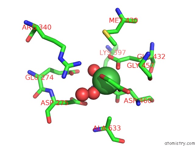
Nickel binding site 1 out of 2 in 5vfb
Go back to
Nickel binding site 1 out
of 2 in the 1.36 Angstrom Resolution Crystal Structure of Malate Synthase G From Pseudomonas Aeruginosa in Complex with Glycolic Acid.
Mono view

Stereo pair view
Mono view
Stereo pair view
A full contact list of Nickel with other atoms in the Ni binding
site number 1 of 1.36 Angstrom Resolution Crystal Structure of Malate Synthase G From Pseudomonas Aeruginosa in Complex with Glycolic Acid. within 5.0Å range:
|
Nickel binding site 2 out of 2 in 5vfb
Go back to
Nickel binding site 2 out
of 2 in the 1.36 Angstrom Resolution Crystal Structure of Malate Synthase G From Pseudomonas Aeruginosa in Complex with Glycolic Acid.

Mono view

Stereo pair view
Mono view
Stereo pair view
A full contact list of Nickel with other atoms in the Ni binding
site number 2 of 1.36 Angstrom Resolution Crystal Structure of Malate Synthase G From Pseudomonas Aeruginosa in Complex with Glycolic Acid. within 5.0Å range:
|
Reference:
G.Minasov,
L.Shuvalova,
I.Dubrovska,
O.Kiryukhina,
S.Grimshaw,
K.Kwon,
W.F.Anderson,
Center For Structural Genomics Of Infectious Diseases(Csgid).
1.36 Angstrom Resolution Crystal Structure of Malate Synthase G From Pseudomonas Aeruginosa in Complex with Glycolic Acid. To Be Published.
Page generated: Thu Oct 10 08:07:35 2024
Last articles
K in 9ES1K in 9EIO
K in 9ED1
K in 9EIE
K in 9EAF
K in 9DWN
K in 9E4V
K in 9DKF
K in 9DTR
K in 9DXX