Nickel »
PDB 5tvr-5xgz »
5x2t »
Nickel in PDB 5x2t: Direct Observation of Conformational Population Shifts in Hemoglobin: Crystal Structure of Half-Liganded Hemoglobin After Adding 4 Mm Bezafibrate pH 7.2.
Protein crystallography data
The structure of Direct Observation of Conformational Population Shifts in Hemoglobin: Crystal Structure of Half-Liganded Hemoglobin After Adding 4 Mm Bezafibrate pH 7.2., PDB code: 5x2t
was solved by
M.Ohki,
S.-Y.Park,
with X-Ray Crystallography technique. A brief refinement statistics is given in the table below:
| Resolution Low / High (Å) | 48.77 / 2.64 |
| Space group | C 1 2 1 |
| Cell size a, b, c (Å), α, β, γ (°) | 228.494, 55.024, 138.504, 90.00, 103.38, 90.00 |
| R / Rfree (%) | 23.2 / 27.7 |
Other elements in 5x2t:
The structure of Direct Observation of Conformational Population Shifts in Hemoglobin: Crystal Structure of Half-Liganded Hemoglobin After Adding 4 Mm Bezafibrate pH 7.2. also contains other interesting chemical elements:
| Iron | (Fe) | 6 atoms |
| Chlorine | (Cl) | 1 atom |
Nickel Binding Sites:
The binding sites of Nickel atom in the Direct Observation of Conformational Population Shifts in Hemoglobin: Crystal Structure of Half-Liganded Hemoglobin After Adding 4 Mm Bezafibrate pH 7.2.
(pdb code 5x2t). This binding sites where shown within
5.0 Angstroms radius around Nickel atom.
In total 6 binding sites of Nickel where determined in the Direct Observation of Conformational Population Shifts in Hemoglobin: Crystal Structure of Half-Liganded Hemoglobin After Adding 4 Mm Bezafibrate pH 7.2., PDB code: 5x2t:
Jump to Nickel binding site number: 1; 2; 3; 4; 5; 6;
In total 6 binding sites of Nickel where determined in the Direct Observation of Conformational Population Shifts in Hemoglobin: Crystal Structure of Half-Liganded Hemoglobin After Adding 4 Mm Bezafibrate pH 7.2., PDB code: 5x2t:
Jump to Nickel binding site number: 1; 2; 3; 4; 5; 6;
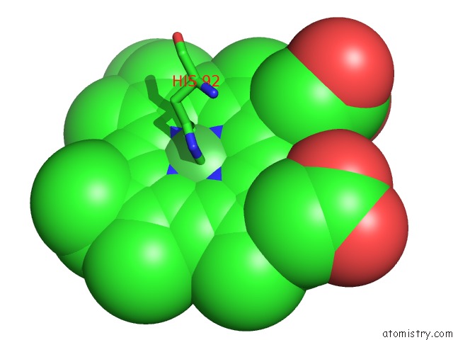
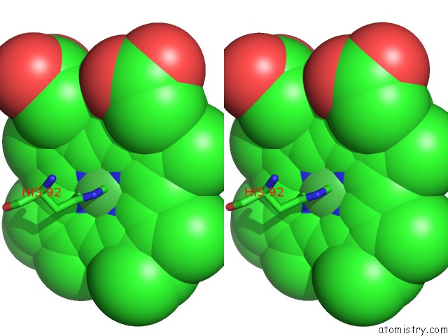
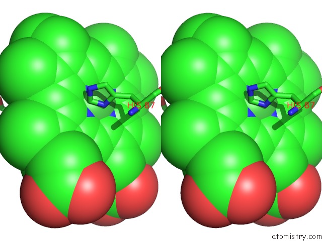
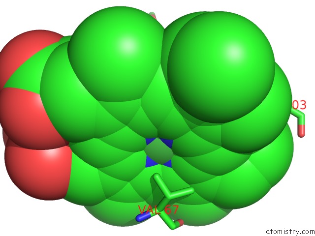
Nickel binding site 1 out of 6 in 5x2t
Go back to
Nickel binding site 1 out
of 6 in the Direct Observation of Conformational Population Shifts in Hemoglobin: Crystal Structure of Half-Liganded Hemoglobin After Adding 4 Mm Bezafibrate pH 7.2.

Mono view

Stereo pair view
Mono view
Stereo pair view
A full contact list of Nickel with other atoms in the Ni binding
site number 1 of Direct Observation of Conformational Population Shifts in Hemoglobin: Crystal Structure of Half-Liganded Hemoglobin After Adding 4 Mm Bezafibrate pH 7.2. within 5.0Å range:
|
Nickel binding site 2 out of 6 in 5x2t
Go back to
Nickel binding site 2 out
of 6 in the Direct Observation of Conformational Population Shifts in Hemoglobin: Crystal Structure of Half-Liganded Hemoglobin After Adding 4 Mm Bezafibrate pH 7.2.
Mono view
Stereo pair view
Mono view
Stereo pair view
A full contact list of Nickel with other atoms in the Ni binding
site number 2 of Direct Observation of Conformational Population Shifts in Hemoglobin: Crystal Structure of Half-Liganded Hemoglobin After Adding 4 Mm Bezafibrate pH 7.2. within 5.0Å range:
|
Nickel binding site 3 out of 6 in 5x2t
Go back to
Nickel binding site 3 out
of 6 in the Direct Observation of Conformational Population Shifts in Hemoglobin: Crystal Structure of Half-Liganded Hemoglobin After Adding 4 Mm Bezafibrate pH 7.2.

Mono view

Stereo pair view
Mono view
Stereo pair view
A full contact list of Nickel with other atoms in the Ni binding
site number 3 of Direct Observation of Conformational Population Shifts in Hemoglobin: Crystal Structure of Half-Liganded Hemoglobin After Adding 4 Mm Bezafibrate pH 7.2. within 5.0Å range:
|
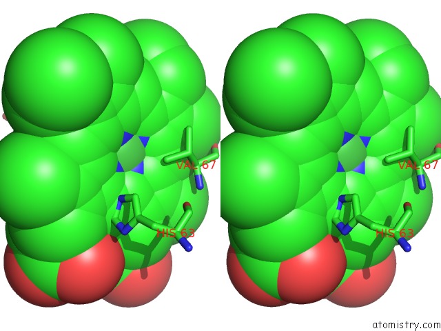
Nickel binding site 4 out of 6 in 5x2t
Go back to
Nickel binding site 4 out
of 6 in the Direct Observation of Conformational Population Shifts in Hemoglobin: Crystal Structure of Half-Liganded Hemoglobin After Adding 4 Mm Bezafibrate pH 7.2.

Mono view
Stereo pair view
Mono view
Stereo pair view
A full contact list of Nickel with other atoms in the Ni binding
site number 4 of Direct Observation of Conformational Population Shifts in Hemoglobin: Crystal Structure of Half-Liganded Hemoglobin After Adding 4 Mm Bezafibrate pH 7.2. within 5.0Å range:
|
Nickel binding site 5 out of 6 in 5x2t
Go back to
Nickel binding site 5 out
of 6 in the Direct Observation of Conformational Population Shifts in Hemoglobin: Crystal Structure of Half-Liganded Hemoglobin After Adding 4 Mm Bezafibrate pH 7.2.
Mono view
Stereo pair view
Mono view
Stereo pair view
A full contact list of Nickel with other atoms in the Ni binding
site number 5 of Direct Observation of Conformational Population Shifts in Hemoglobin: Crystal Structure of Half-Liganded Hemoglobin After Adding 4 Mm Bezafibrate pH 7.2. within 5.0Å range:
|
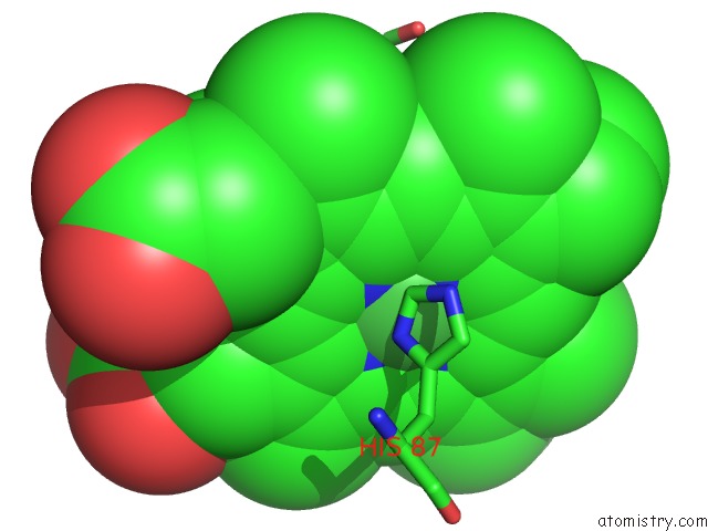
Nickel binding site 6 out of 6 in 5x2t
Go back to
Nickel binding site 6 out
of 6 in the Direct Observation of Conformational Population Shifts in Hemoglobin: Crystal Structure of Half-Liganded Hemoglobin After Adding 4 Mm Bezafibrate pH 7.2.
Mono view

Stereo pair view
Mono view
Stereo pair view
A full contact list of Nickel with other atoms in the Ni binding
site number 6 of Direct Observation of Conformational Population Shifts in Hemoglobin: Crystal Structure of Half-Liganded Hemoglobin After Adding 4 Mm Bezafibrate pH 7.2. within 5.0Å range:
|
Reference:
N.Shibayama,
M.Ohki,
J.R.H.Tame,
S.Y.Park.
Direct Observation of Conformational Population Shifts in Crystalline Human Hemoglobin. J. Biol. Chem. V. 292 18258 2017.
ISSN: ESSN 1083-351X
PubMed: 28931607
DOI: 10.1074/JBC.M117.781146
Page generated: Thu Oct 10 08:10:29 2024
ISSN: ESSN 1083-351X
PubMed: 28931607
DOI: 10.1074/JBC.M117.781146
Last articles
Mg in 3I5XMg in 3I4K
Mg in 3I5F
Mg in 3I5C
Mg in 3I4N
Mg in 3I3E
Mg in 3I4D
Mg in 3I4M
Mg in 3I3D
Mg in 3I3B