Nickel »
PDB 6cac-6f5r »
6ec3 »
Nickel in PDB 6ec3: Crystal Structure of EVDMO1
Protein crystallography data
The structure of Crystal Structure of EVDMO1, PDB code: 6ec3
was solved by
K.M.Mcculloch,
T.M.Iverson,
C.A.Starbird,
N.A.Perry,
Q.Chen,
S.Berndt,
I.Yamakawa,
L.V.Loukachevitch,
with X-Ray Crystallography technique. A brief refinement statistics is given in the table below:
| Resolution Low / High (Å) | 45.64 / 3.35 |
| Space group | I 41 2 2 |
| Cell size a, b, c (Å), α, β, γ (°) | 191.633, 191.633, 269.618, 90.00, 90.00, 90.00 |
| R / Rfree (%) | 22.1 / 26.6 |
Nickel Binding Sites:
The binding sites of Nickel atom in the Crystal Structure of EVDMO1
(pdb code 6ec3). This binding sites where shown within
5.0 Angstroms radius around Nickel atom.
In total 3 binding sites of Nickel where determined in the Crystal Structure of EVDMO1, PDB code: 6ec3:
Jump to Nickel binding site number: 1; 2; 3;
In total 3 binding sites of Nickel where determined in the Crystal Structure of EVDMO1, PDB code: 6ec3:
Jump to Nickel binding site number: 1; 2; 3;
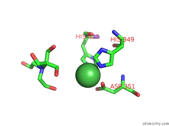
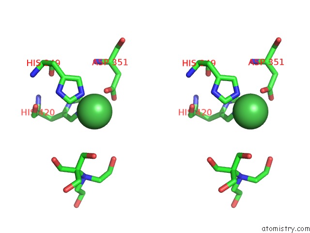
Nickel binding site 1 out of 3 in 6ec3
Go back to
Nickel binding site 1 out
of 3 in the Crystal Structure of EVDMO1
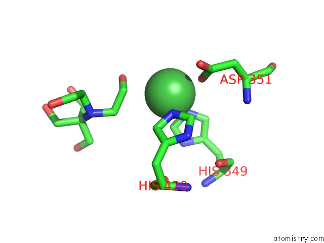
Mono view
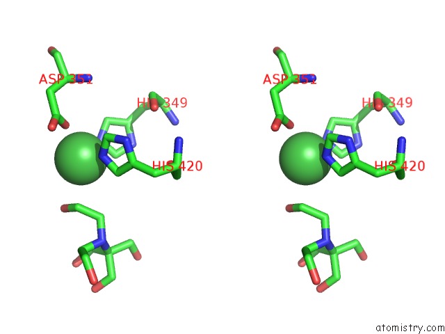
Stereo pair view
Mono view
Stereo pair view
A full contact list of Nickel with other atoms in the Ni binding
site number 1 of Crystal Structure of EVDMO1 within 5.0Å range:
|
Nickel binding site 2 out of 3 in 6ec3
Go back to
Nickel binding site 2 out
of 3 in the Crystal Structure of EVDMO1
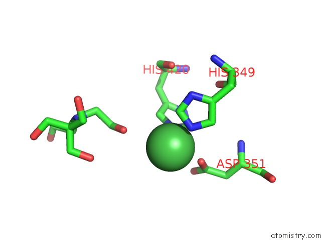
Mono view
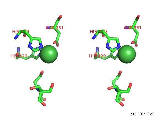
Stereo pair view
Mono view
Stereo pair view
A full contact list of Nickel with other atoms in the Ni binding
site number 2 of Crystal Structure of EVDMO1 within 5.0Å range:
|
Nickel binding site 3 out of 3 in 6ec3
Go back to
Nickel binding site 3 out
of 3 in the Crystal Structure of EVDMO1
Mono view
Stereo pair view
Mono view
Stereo pair view
A full contact list of Nickel with other atoms in the Ni binding
site number 3 of Crystal Structure of EVDMO1 within 5.0Å range:
|
Reference:
C.A.Starbird,
N.A.Perry,
Q.Chen,
S.Berndt,
I.Yamakawa,
L.V.Loukachevitch,
E.M.Limbrick,
B.O.Bachmann,
T.M.Iverson,
K.M.Mcculloch.
The Structure of the Bifunctional Everninomicin Biosynthetic Enzyme EVDMO1 Suggests Independent Activity of the Fused Methyltransferase-Oxidase Domains. Biochemistry V. 57 6827 2018.
ISSN: ISSN 1520-4995
PubMed: 30525509
DOI: 10.1021/ACS.BIOCHEM.8B00836
Page generated: Thu Oct 10 08:23:25 2024
ISSN: ISSN 1520-4995
PubMed: 30525509
DOI: 10.1021/ACS.BIOCHEM.8B00836
Last articles
Mg in 3J7HMg in 3J7I
Mg in 3IVK
Mg in 3J6E
Mg in 3J6G
Mg in 3J6P
Mg in 3J6H
Mg in 3J1F
Mg in 3J6F
Mg in 3J5V