Nickel »
PDB 6ikt-6lrx »
6jfs »
Nickel in PDB 6jfs: K4U Bound Crystal Structure of Class II Peptide Deformylase From Methicillin Resistant Staphylococcus Aureus
Enzymatic activity of K4U Bound Crystal Structure of Class II Peptide Deformylase From Methicillin Resistant Staphylococcus Aureus
All present enzymatic activity of K4U Bound Crystal Structure of Class II Peptide Deformylase From Methicillin Resistant Staphylococcus Aureus:
3.5.1.88;
3.5.1.88;
Protein crystallography data
The structure of K4U Bound Crystal Structure of Class II Peptide Deformylase From Methicillin Resistant Staphylococcus Aureus, PDB code: 6jfs
was solved by
I.H.Lee,
T.H.Ho,
L.W.Kang,
with X-Ray Crystallography technique. A brief refinement statistics is given in the table below:
| Resolution Low / High (Å) | 33.64 / 2.25 |
| Space group | C 2 2 21 |
| Cell size a, b, c (Å), α, β, γ (°) | 95.760, 120.396, 47.208, 90.00, 90.00, 90.00 |
| R / Rfree (%) | 20.9 / 26.8 |
Nickel Binding Sites:
The binding sites of Nickel atom in the K4U Bound Crystal Structure of Class II Peptide Deformylase From Methicillin Resistant Staphylococcus Aureus
(pdb code 6jfs). This binding sites where shown within
5.0 Angstroms radius around Nickel atom.
In total only one binding site of Nickel was determined in the K4U Bound Crystal Structure of Class II Peptide Deformylase From Methicillin Resistant Staphylococcus Aureus, PDB code: 6jfs:
In total only one binding site of Nickel was determined in the K4U Bound Crystal Structure of Class II Peptide Deformylase From Methicillin Resistant Staphylococcus Aureus, PDB code: 6jfs:
Nickel binding site 1 out of 1 in 6jfs
Go back to
Nickel binding site 1 out
of 1 in the K4U Bound Crystal Structure of Class II Peptide Deformylase From Methicillin Resistant Staphylococcus Aureus
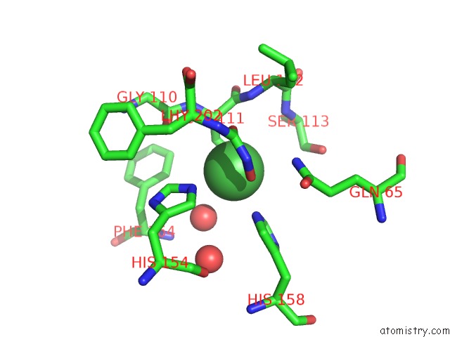
Mono view
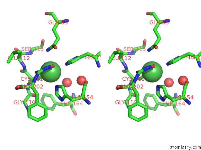
Stereo pair view
Mono view
Stereo pair view
A full contact list of Nickel with other atoms in the Ni binding
site number 1 of K4U Bound Crystal Structure of Class II Peptide Deformylase From Methicillin Resistant Staphylococcus Aureus within 5.0Å range:
|
Reference:
I.H.Lee,
T.H.Ho,
L.W.Kang.
K4U Bound Crystal Structure of Class II Peptide Deformylase From Methicillin Resistant Staphylococcus Aureus To Be Published.
Page generated: Thu Oct 10 08:34:49 2024
Last articles
Mg in 5EXAMg in 5EWU
Mg in 5EX5
Mg in 5EX2
Mg in 5EX1
Mg in 5EW7
Mg in 5EW4
Mg in 5EVZ
Mg in 5ETS
Mg in 5ETT