Nickel »
PDB 6r44-6vwy »
6vfu »
Nickel in PDB 6vfu: Crystal Structure of Human Protocadherin 19 EC1-EC4
Protein crystallography data
The structure of Crystal Structure of Human Protocadherin 19 EC1-EC4, PDB code: 6vfu
was solved by
O.J.Harrison,
J.Brasch,
L.Shapiro,
with X-Ray Crystallography technique. A brief refinement statistics is given in the table below:
| Resolution Low / High (Å) | 19.99 / 3.50 |
| Space group | P 32 2 1 |
| Cell size a, b, c (Å), α, β, γ (°) | 108.971, 108.971, 309.663, 90.00, 90.00, 120.00 |
| R / Rfree (%) | 26.5 / 29.2 |
Other elements in 6vfu:
The structure of Crystal Structure of Human Protocadherin 19 EC1-EC4 also contains other interesting chemical elements:
| Calcium | (Ca) | 27 atoms |
| Chlorine | (Cl) | 4 atoms |
Nickel Binding Sites:
The binding sites of Nickel atom in the Crystal Structure of Human Protocadherin 19 EC1-EC4
(pdb code 6vfu). This binding sites where shown within
5.0 Angstroms radius around Nickel atom.
In total 2 binding sites of Nickel where determined in the Crystal Structure of Human Protocadherin 19 EC1-EC4, PDB code: 6vfu:
Jump to Nickel binding site number: 1; 2;
In total 2 binding sites of Nickel where determined in the Crystal Structure of Human Protocadherin 19 EC1-EC4, PDB code: 6vfu:
Jump to Nickel binding site number: 1; 2;
Nickel binding site 1 out of 2 in 6vfu
Go back to
Nickel binding site 1 out
of 2 in the Crystal Structure of Human Protocadherin 19 EC1-EC4
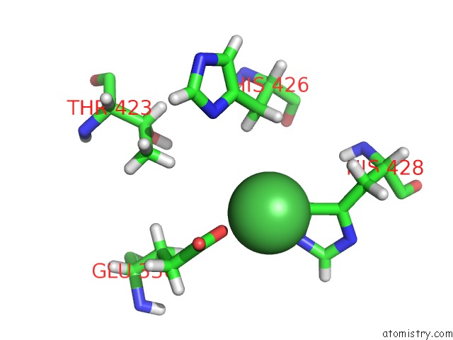
Mono view
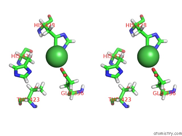
Stereo pair view
Mono view
Stereo pair view
A full contact list of Nickel with other atoms in the Ni binding
site number 1 of Crystal Structure of Human Protocadherin 19 EC1-EC4 within 5.0Å range:
|
Nickel binding site 2 out of 2 in 6vfu
Go back to
Nickel binding site 2 out
of 2 in the Crystal Structure of Human Protocadherin 19 EC1-EC4
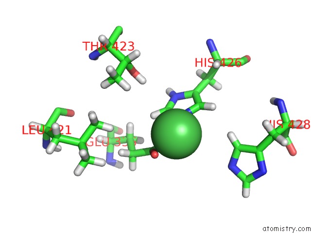
Mono view
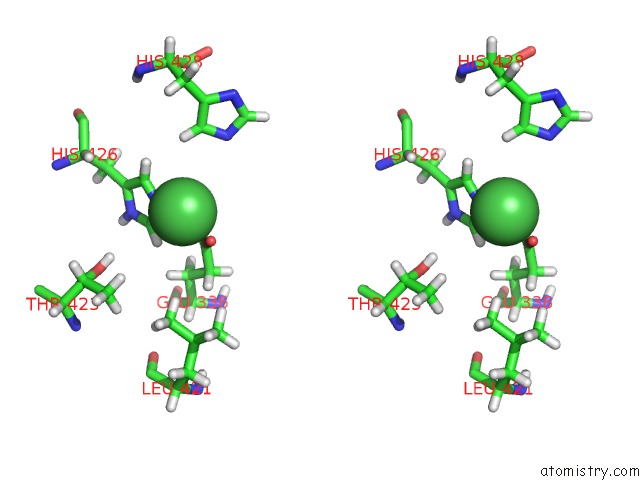
Stereo pair view
Mono view
Stereo pair view
A full contact list of Nickel with other atoms in the Ni binding
site number 2 of Crystal Structure of Human Protocadherin 19 EC1-EC4 within 5.0Å range:
|
Reference:
O.J.Harrison,
J.Brasch,
P.S.Katsamba,
G.Ahlsen,
A.J.Noble,
H.Dan,
R.V.Sampogna,
C.S.Potter,
B.Carragher,
B.Honig,
L.Shapiro.
Family-Wide Structural and Biophysical Analysis of Binding Interactions Among Non-Clustered Delta-Protocadherins. Cell Rep V. 30 2655 2020.
ISSN: ESSN 2211-1247
PubMed: 32101743
DOI: 10.1016/J.CELREP.2020.02.003
Page generated: Thu Oct 10 08:55:18 2024
ISSN: ESSN 2211-1247
PubMed: 32101743
DOI: 10.1016/J.CELREP.2020.02.003
Last articles
Fe in 9O8UFe in 9N5V
Fe in 9NSE
Fe in 9NQU
Fe in 9NP6
Fe in 9NH3
Fe in 9NEA
Fe in 9NE9
Fe in 9NE8
Fe in 9NE7