Nickel »
PDB 6vwz-6z8m »
6wwx »
Nickel in PDB 6wwx: Crystal Structure of Truncated Bacteriophage Hyaluronan Lyase Hylp in Complex with Unsaturated Hyaluronan Tetra-Saccharides
Enzymatic activity of Crystal Structure of Truncated Bacteriophage Hyaluronan Lyase Hylp in Complex with Unsaturated Hyaluronan Tetra-Saccharides
All present enzymatic activity of Crystal Structure of Truncated Bacteriophage Hyaluronan Lyase Hylp in Complex with Unsaturated Hyaluronan Tetra-Saccharides:
3.2.1.35;
3.2.1.35;
Protein crystallography data
The structure of Crystal Structure of Truncated Bacteriophage Hyaluronan Lyase Hylp in Complex with Unsaturated Hyaluronan Tetra-Saccharides, PDB code: 6wwx
was solved by
C.Deivanayagam,
N.Schormann,
with X-Ray Crystallography technique. A brief refinement statistics is given in the table below:
| Resolution Low / High (Å) | 38.68 / 2.20 |
| Space group | P 21 21 21 |
| Cell size a, b, c (Å), α, β, γ (°) | 67.535, 86.819, 168.646, 90, 90, 90 |
| R / Rfree (%) | 18.2 / 21 |
Nickel Binding Sites:
The binding sites of Nickel atom in the Crystal Structure of Truncated Bacteriophage Hyaluronan Lyase Hylp in Complex with Unsaturated Hyaluronan Tetra-Saccharides
(pdb code 6wwx). This binding sites where shown within
5.0 Angstroms radius around Nickel atom.
In total only one binding site of Nickel was determined in the Crystal Structure of Truncated Bacteriophage Hyaluronan Lyase Hylp in Complex with Unsaturated Hyaluronan Tetra-Saccharides, PDB code: 6wwx:
In total only one binding site of Nickel was determined in the Crystal Structure of Truncated Bacteriophage Hyaluronan Lyase Hylp in Complex with Unsaturated Hyaluronan Tetra-Saccharides, PDB code: 6wwx:
Nickel binding site 1 out of 1 in 6wwx
Go back to
Nickel binding site 1 out
of 1 in the Crystal Structure of Truncated Bacteriophage Hyaluronan Lyase Hylp in Complex with Unsaturated Hyaluronan Tetra-Saccharides
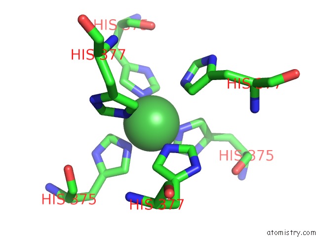
Mono view
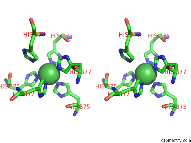
Stereo pair view
Mono view
Stereo pair view
A full contact list of Nickel with other atoms in the Ni binding
site number 1 of Crystal Structure of Truncated Bacteriophage Hyaluronan Lyase Hylp in Complex with Unsaturated Hyaluronan Tetra-Saccharides within 5.0Å range:
|
Reference:
J.H.Lee,
N.Schormann,
K.R.Rajashankar,
C.Deivanaygam.
Crystal Structure of Streptococcal Bacteriophage Hyaluronidase: Presence of A Prokaryotic Collagen and Elucidation of Catalytic Mechanism To Be Published.
Page generated: Mon Aug 18 21:36:31 2025
Last articles
Pt in 7BPRPt in 6YK8
Pt in 6ZRW
Pt in 7BD7
Pt in 6ZZ9
Pt in 6XCL
Pt in 6ZLX
Pt in 6SWJ
Pt in 6MYL
Pt in 6WX5