Nickel »
PDB 6vwz-6z8m »
6xnw »
Nickel in PDB 6xnw: Crystal Structure of V39A Mutant of Human CEACAM1
Protein crystallography data
The structure of Crystal Structure of V39A Mutant of Human CEACAM1, PDB code: 6xnw
was solved by
A.K.Gandhi,
W.M.Kim,
Z.-Y.Sun,
Y.H.Huang,
D.Bonsor,
G.A.Petsko,
V.Kuchroo,
R.S.Blumberg,
with X-Ray Crystallography technique. A brief refinement statistics is given in the table below:
| Resolution Low / High (Å) | 39.59 / 1.90 |
| Space group | P 3 |
| Cell size a, b, c (Å), α, β, γ (°) | 91.44, 91.44, 64.41, 90, 90, 120 |
| R / Rfree (%) | 14.5 / 18.6 |
Nickel Binding Sites:
The binding sites of Nickel atom in the Crystal Structure of V39A Mutant of Human CEACAM1
(pdb code 6xnw). This binding sites where shown within
5.0 Angstroms radius around Nickel atom.
In total 2 binding sites of Nickel where determined in the Crystal Structure of V39A Mutant of Human CEACAM1, PDB code: 6xnw:
Jump to Nickel binding site number: 1; 2;
In total 2 binding sites of Nickel where determined in the Crystal Structure of V39A Mutant of Human CEACAM1, PDB code: 6xnw:
Jump to Nickel binding site number: 1; 2;
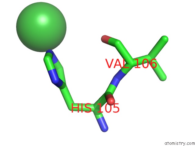
Nickel binding site 1 out of 2 in 6xnw
Go back to
Nickel binding site 1 out
of 2 in the Crystal Structure of V39A Mutant of Human CEACAM1

Mono view
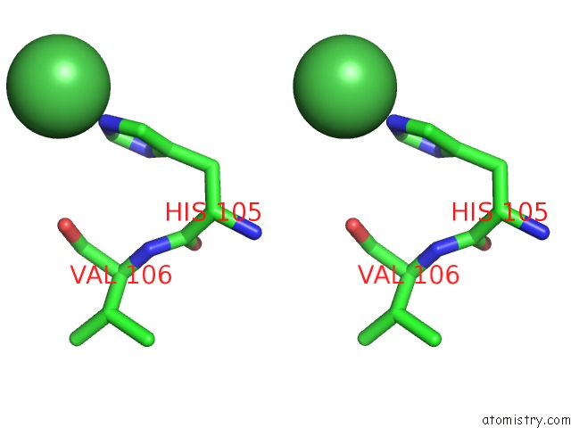
Stereo pair view
Mono view
Stereo pair view
A full contact list of Nickel with other atoms in the Ni binding
site number 1 of Crystal Structure of V39A Mutant of Human CEACAM1 within 5.0Å range:
|
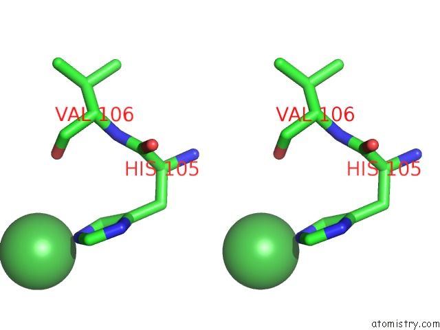
Nickel binding site 2 out of 2 in 6xnw
Go back to
Nickel binding site 2 out
of 2 in the Crystal Structure of V39A Mutant of Human CEACAM1
Mono view
Stereo pair view
Mono view
Stereo pair view
A full contact list of Nickel with other atoms in the Ni binding
site number 2 of Crystal Structure of V39A Mutant of Human CEACAM1 within 5.0Å range:
|
Reference:
A.K.Gandhi,
Z.J.Sun,
W.M.Kim,
Y.H.Huang,
D.A.Bonsor,
E.J.Sundberg,
Y.Kondo,
G.Wagner,
V.K.Kuchroo,
G.Petsko,
R.S.Blumberg.
Structural Basis of the Dynamic Human CEACAM1 Monomer-Dimer Equilibrium Commun Biol 2021.
ISSN: ESSN 2399-3642
DOI: 10.1038/S42003-021-01871-2
Page generated: Thu Oct 10 08:58:27 2024
ISSN: ESSN 2399-3642
DOI: 10.1038/S42003-021-01871-2
Last articles
Fe in 2YXOFe in 2YRS
Fe in 2YXC
Fe in 2YNM
Fe in 2YVJ
Fe in 2YP1
Fe in 2YU2
Fe in 2YU1
Fe in 2YQB
Fe in 2YOO