Nickel »
PDB 7odh-7utd »
7u0v »
Nickel in PDB 7u0v: Structure of Myxoma Virus M062 Protein Variant Lau
Protein crystallography data
The structure of Structure of Myxoma Virus M062 Protein Variant Lau, PDB code: 7u0v
was solved by
P.O'byrne,
A.R.Khan,
with X-Ray Crystallography technique. A brief refinement statistics is given in the table below:
| Resolution Low / High (Å) | 47.41 / 2.45 |
| Space group | P 64 |
| Cell size a, b, c (Å), α, β, γ (°) | 94.827, 94.827, 44.136, 90, 90, 120 |
| R / Rfree (%) | 21.1 / 26.6 |
Nickel Binding Sites:
The binding sites of Nickel atom in the Structure of Myxoma Virus M062 Protein Variant Lau
(pdb code 7u0v). This binding sites where shown within
5.0 Angstroms radius around Nickel atom.
In total only one binding site of Nickel was determined in the Structure of Myxoma Virus M062 Protein Variant Lau, PDB code: 7u0v:
In total only one binding site of Nickel was determined in the Structure of Myxoma Virus M062 Protein Variant Lau, PDB code: 7u0v:
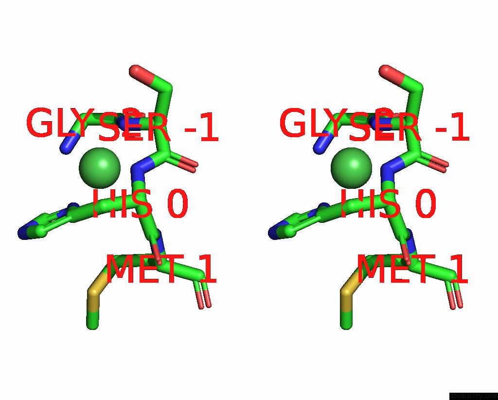
Nickel binding site 1 out of 1 in 7u0v
Go back to
Nickel binding site 1 out
of 1 in the Structure of Myxoma Virus M062 Protein Variant Lau

Mono view
Stereo pair view
Mono view
Stereo pair view
A full contact list of Nickel with other atoms in the Ni binding
site number 1 of Structure of Myxoma Virus M062 Protein Variant Lau within 5.0Å range:
|
Reference:
P.O'byrne,
A.R.Khan.
Structure of Myxoma Virus M062 Protein Variant Mav To Be Published.
Page generated: Thu Oct 10 09:24:56 2024
Last articles
Fe in 2YXOFe in 2YRS
Fe in 2YXC
Fe in 2YNM
Fe in 2YVJ
Fe in 2YP1
Fe in 2YU2
Fe in 2YU1
Fe in 2YQB
Fe in 2YOO