Nickel »
PDB 1jhj-1q0d »
1jmz »
Nickel in PDB 1jmz: Crystal Structure of A Quinohemoprotein Amine Dehydrogenase From Pseudomonas Putida with Inhibitor
Protein crystallography data
The structure of Crystal Structure of A Quinohemoprotein Amine Dehydrogenase From Pseudomonas Putida with Inhibitor, PDB code: 1jmz
was solved by
A.Satoh,
I.Miyahara,
K.Hirotsu,
with X-Ray Crystallography technique. A brief refinement statistics is given in the table below:
| Resolution Low / High (Å) | 20.00 / 2.00 |
| Space group | C 1 2 1 |
| Cell size a, b, c (Å), α, β, γ (°) | 167.210, 92.370, 79.300, 90.00, 112.00, 90.00 |
| R / Rfree (%) | 21.5 / 25.1 |
Other elements in 1jmz:
The structure of Crystal Structure of A Quinohemoprotein Amine Dehydrogenase From Pseudomonas Putida with Inhibitor also contains other interesting chemical elements:
| Iron | (Fe) | 2 atoms |
Nickel Binding Sites:
The binding sites of Nickel atom in the Crystal Structure of A Quinohemoprotein Amine Dehydrogenase From Pseudomonas Putida with Inhibitor
(pdb code 1jmz). This binding sites where shown within
5.0 Angstroms radius around Nickel atom.
In total only one binding site of Nickel was determined in the Crystal Structure of A Quinohemoprotein Amine Dehydrogenase From Pseudomonas Putida with Inhibitor, PDB code: 1jmz:
In total only one binding site of Nickel was determined in the Crystal Structure of A Quinohemoprotein Amine Dehydrogenase From Pseudomonas Putida with Inhibitor, PDB code: 1jmz:
Nickel binding site 1 out of 1 in 1jmz
Go back to
Nickel binding site 1 out
of 1 in the Crystal Structure of A Quinohemoprotein Amine Dehydrogenase From Pseudomonas Putida with Inhibitor
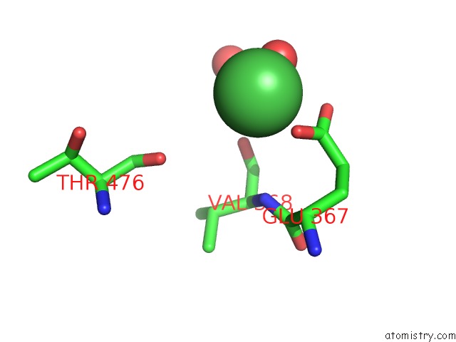
Mono view
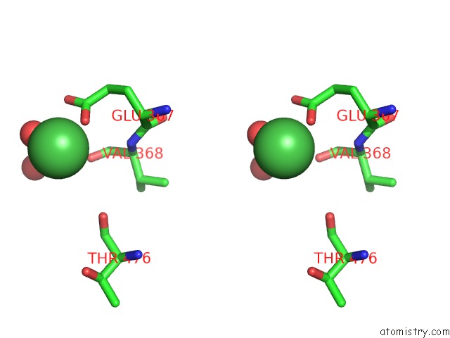
Stereo pair view
Mono view
Stereo pair view
A full contact list of Nickel with other atoms in the Ni binding
site number 1 of Crystal Structure of A Quinohemoprotein Amine Dehydrogenase From Pseudomonas Putida with Inhibitor within 5.0Å range:
|
Reference:
A.Satoh,
J.K.Kim,
I.Miyahara,
B.Devreese,
I.Vandenberghe,
A.Hacisalihoglu,
T.Okajima,
S.Kuroda,
O.Adachi,
J.A.Duine,
J.Van Beeumen,
K.Tanizawa,
K.Hirotsu.
Crystal Structure of Quinohemoprotein Amine Dehydrogenase From Pseudomonas Putida. Identification of A Novel Quinone Cofactor Encaged By Multiple Thioether Cross-Bridges. J.Biol.Chem. V. 277 2830 2002.
ISSN: ISSN 0021-9258
PubMed: 11704672
DOI: 10.1074/JBC.M109090200
Page generated: Wed Oct 9 15:00:42 2024
ISSN: ISSN 0021-9258
PubMed: 11704672
DOI: 10.1074/JBC.M109090200
Last articles
Zn in 9MJ5Zn in 9HNW
Zn in 9G0L
Zn in 9FNE
Zn in 9DZN
Zn in 9E0I
Zn in 9D32
Zn in 9DAK
Zn in 8ZXC
Zn in 8ZUF