Nickel »
PDB 1jhj-1q0d »
1k2e »
Nickel in PDB 1k2e: Crystal Structure of A Nudix Protein From Pyrobaculum Aerophilum
Protein crystallography data
The structure of Crystal Structure of A Nudix Protein From Pyrobaculum Aerophilum, PDB code: 1k2e
was solved by
S.Wang,
C.Mura,
M.R.Sawaya,
D.Cascio,
D.Eisenberg,
with X-Ray Crystallography technique. A brief refinement statistics is given in the table below:
| Resolution Low / High (Å) | 19.74 / 1.80 |
| Space group | P 21 21 21 |
| Cell size a, b, c (Å), α, β, γ (°) | 52.712, 72.612, 85.175, 90.00, 90.00, 90.00 |
| R / Rfree (%) | 18.4 / 22 |
Nickel Binding Sites:
The binding sites of Nickel atom in the Crystal Structure of A Nudix Protein From Pyrobaculum Aerophilum
(pdb code 1k2e). This binding sites where shown within
5.0 Angstroms radius around Nickel atom.
In total 2 binding sites of Nickel where determined in the Crystal Structure of A Nudix Protein From Pyrobaculum Aerophilum, PDB code: 1k2e:
Jump to Nickel binding site number: 1; 2;
In total 2 binding sites of Nickel where determined in the Crystal Structure of A Nudix Protein From Pyrobaculum Aerophilum, PDB code: 1k2e:
Jump to Nickel binding site number: 1; 2;
Nickel binding site 1 out of 2 in 1k2e
Go back to
Nickel binding site 1 out
of 2 in the Crystal Structure of A Nudix Protein From Pyrobaculum Aerophilum
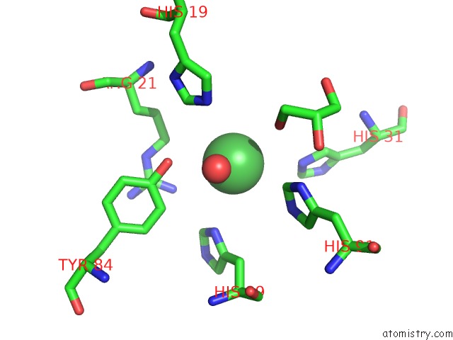
Mono view
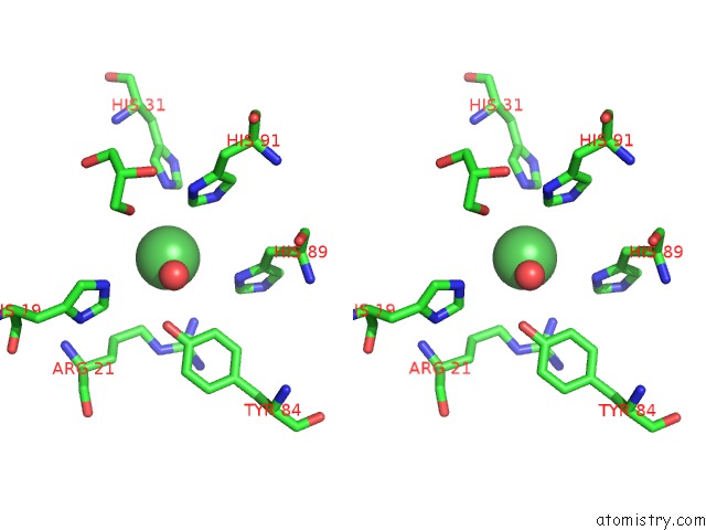
Stereo pair view
Mono view
Stereo pair view
A full contact list of Nickel with other atoms in the Ni binding
site number 1 of Crystal Structure of A Nudix Protein From Pyrobaculum Aerophilum within 5.0Å range:
|
Nickel binding site 2 out of 2 in 1k2e
Go back to
Nickel binding site 2 out
of 2 in the Crystal Structure of A Nudix Protein From Pyrobaculum Aerophilum
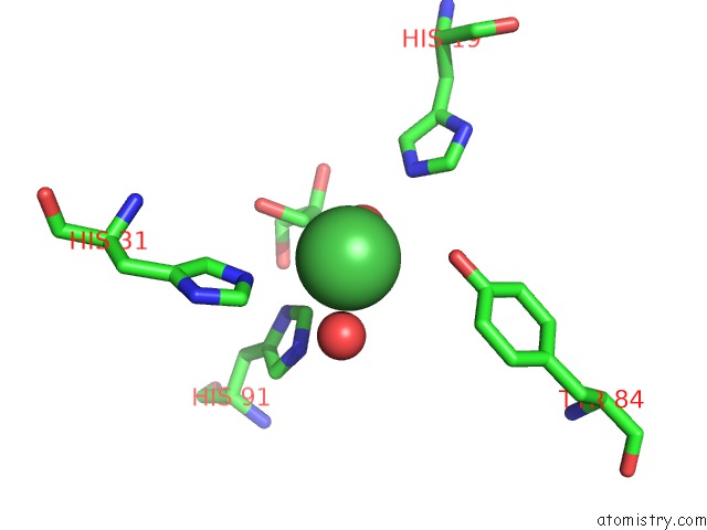
Mono view
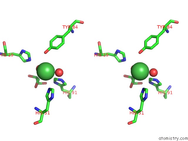
Stereo pair view
Mono view
Stereo pair view
A full contact list of Nickel with other atoms in the Ni binding
site number 2 of Crystal Structure of A Nudix Protein From Pyrobaculum Aerophilum within 5.0Å range:
|
Reference:
S.Wang,
C.Mura,
M.R.Sawaya,
D.Cascio,
D.Eisenberg.
Structure of A Nudix Protein From Pyrobaculum Aerophilum Reveals A Dimer with Two Intersubunit Beta-Sheets. Acta Crystallogr.,Sect.D V. 58 571 2002.
ISSN: ISSN 0907-4449
PubMed: 11914479
DOI: 10.1107/S0907444902001191
Page generated: Wed Oct 9 15:00:55 2024
ISSN: ISSN 0907-4449
PubMed: 11914479
DOI: 10.1107/S0907444902001191
Last articles
Zn in 9MJ5Zn in 9HNW
Zn in 9G0L
Zn in 9FNE
Zn in 9DZN
Zn in 9E0I
Zn in 9D32
Zn in 9DAK
Zn in 8ZXC
Zn in 8ZUF