Nickel »
PDB 3a1f-3ds9 »
3bix »
Nickel in PDB 3bix: Crystal Structure of the Extracellular Esterase Domain of Neuroligin-1
Protein crystallography data
The structure of Crystal Structure of the Extracellular Esterase Domain of Neuroligin-1, PDB code: 3bix
was solved by
D.Arac,
A.A.Boucard,
E.Ozkan,
P.Strop,
E.Newell,
T.C.Sudhof,
A.T.Brunger,
with X-Ray Crystallography technique. A brief refinement statistics is given in the table below:
| Resolution Low / High (Å) | 45.88 / 1.80 |
| Space group | P 32 |
| Cell size a, b, c (Å), α, β, γ (°) | 173.744, 173.744, 108.045, 90.00, 90.00, 120.00 |
| R / Rfree (%) | 18.5 / 20.5 |
Nickel Binding Sites:
The binding sites of Nickel atom in the Crystal Structure of the Extracellular Esterase Domain of Neuroligin-1
(pdb code 3bix). This binding sites where shown within
5.0 Angstroms radius around Nickel atom.
In total 2 binding sites of Nickel where determined in the Crystal Structure of the Extracellular Esterase Domain of Neuroligin-1, PDB code: 3bix:
Jump to Nickel binding site number: 1; 2;
In total 2 binding sites of Nickel where determined in the Crystal Structure of the Extracellular Esterase Domain of Neuroligin-1, PDB code: 3bix:
Jump to Nickel binding site number: 1; 2;
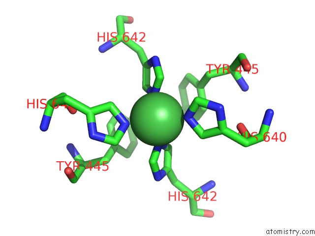
Nickel binding site 1 out of 2 in 3bix
Go back to
Nickel binding site 1 out
of 2 in the Crystal Structure of the Extracellular Esterase Domain of Neuroligin-1

Mono view

Stereo pair view
Mono view
Stereo pair view
A full contact list of Nickel with other atoms in the Ni binding
site number 1 of Crystal Structure of the Extracellular Esterase Domain of Neuroligin-1 within 5.0Å range:
|
Nickel binding site 2 out of 2 in 3bix
Go back to
Nickel binding site 2 out
of 2 in the Crystal Structure of the Extracellular Esterase Domain of Neuroligin-1
Mono view

Stereo pair view
Mono view
Stereo pair view
A full contact list of Nickel with other atoms in the Ni binding
site number 2 of Crystal Structure of the Extracellular Esterase Domain of Neuroligin-1 within 5.0Å range:
|
Reference:
D.Arac,
A.A.Boucard,
E.Ozkan,
P.Strop,
E.Newell,
T.C.Sudhof,
A.T.Brunger.
Structures of Neuroligin-1 and the Neuroligin-1/Neurexin-1BETA Complex Reveal Specific Protein-Protein and Protein-Ca(2+) Interactions. Neuron V. 56 992 2007.
ISSN: ISSN 0896-6273
PubMed: 18093522
DOI: 10.1016/J.NEURON.2007.12.002
Page generated: Wed Oct 9 17:12:19 2024
ISSN: ISSN 0896-6273
PubMed: 18093522
DOI: 10.1016/J.NEURON.2007.12.002
Last articles
Zn in 9MJ5Zn in 9HNW
Zn in 9G0L
Zn in 9FNE
Zn in 9DZN
Zn in 9E0I
Zn in 9D32
Zn in 9DAK
Zn in 8ZXC
Zn in 8ZUF