Nickel »
PDB 3dse-3hy3 »
3gc8 »
Nickel in PDB 3gc8: The Structure of P38BETA C162S in Complex with A Dihydroquinazolinone
Enzymatic activity of The Structure of P38BETA C162S in Complex with A Dihydroquinazolinone
All present enzymatic activity of The Structure of P38BETA C162S in Complex with A Dihydroquinazolinone:
2.7.11.24;
2.7.11.24;
Protein crystallography data
The structure of The Structure of P38BETA C162S in Complex with A Dihydroquinazolinone, PDB code: 3gc8
was solved by
G.Scapin,
S.B.Patel,
with X-Ray Crystallography technique. A brief refinement statistics is given in the table below:
| Resolution Low / High (Å) | 30.00 / 2.40 |
| Space group | P 1 21 1 |
| Cell size a, b, c (Å), α, β, γ (°) | 39.124, 157.710, 60.360, 90.00, 91.57, 90.00 |
| R / Rfree (%) | 20.8 / 27.2 |
Other elements in 3gc8:
The structure of The Structure of P38BETA C162S in Complex with A Dihydroquinazolinone also contains other interesting chemical elements:
| Fluorine | (F) | 2 atoms |
| Chlorine | (Cl) | 6 atoms |
| Sodium | (Na) | 2 atoms |
Nickel Binding Sites:
The binding sites of Nickel atom in the The Structure of P38BETA C162S in Complex with A Dihydroquinazolinone
(pdb code 3gc8). This binding sites where shown within
5.0 Angstroms radius around Nickel atom.
In total only one binding site of Nickel was determined in the The Structure of P38BETA C162S in Complex with A Dihydroquinazolinone, PDB code: 3gc8:
In total only one binding site of Nickel was determined in the The Structure of P38BETA C162S in Complex with A Dihydroquinazolinone, PDB code: 3gc8:
Nickel binding site 1 out of 1 in 3gc8
Go back to
Nickel binding site 1 out
of 1 in the The Structure of P38BETA C162S in Complex with A Dihydroquinazolinone
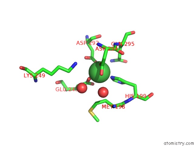
Mono view
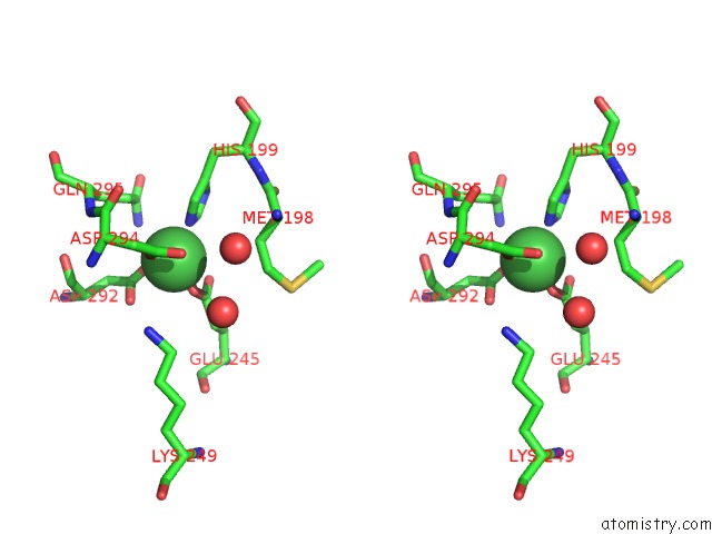
Stereo pair view
Mono view
Stereo pair view
A full contact list of Nickel with other atoms in the Ni binding
site number 1 of The Structure of P38BETA C162S in Complex with A Dihydroquinazolinone within 5.0Å range:
|
Reference:
S.B.Patel,
P.M.Cameron,
S.J.O'keefe,
B.Frantz-Wattley,
J.Thompson,
E.A.O'neill,
T.Tennis,
L.Liu,
J.W.Becker,
G.Scapin.
The Three-Dimensional Structure of Map Kinase P38BETA: Different Features of the Atp-Binding Site in P38BETA Compared with P38ALPHA. Acta Crystallogr.,Sect.D V. 65 777 2009.
ISSN: ISSN 0907-4449
PubMed: 19622861
DOI: 10.1107/S090744490901600X
Page generated: Mon Aug 18 18:37:44 2025
ISSN: ISSN 0907-4449
PubMed: 19622861
DOI: 10.1107/S090744490901600X
Last articles
Ni in 5X2TNi in 5X2R
Ni in 5X2S
Ni in 5WTQ
Ni in 5WXK
Ni in 5W1F
Ni in 5WWX
Ni in 5WRK
Ni in 5WK0
Ni in 5WAY