Nickel »
PDB 3hy4-3kvb »
3i0o »
Nickel in PDB 3i0o: Crystal Structure of Spectinomycin Phosphotransferase, Aph(9)-Ia, in Complex with Adp and Spectinomcyin
Protein crystallography data
The structure of Crystal Structure of Spectinomycin Phosphotransferase, Aph(9)-Ia, in Complex with Adp and Spectinomcyin, PDB code: 3i0o
was solved by
D.H.Fong,
C.T.Lemke,
J.Hwang,
B.Xiong,
A.M.Berghuis,
with X-Ray Crystallography technique. A brief refinement statistics is given in the table below:
| Resolution Low / High (Å) | 37.89 / 2.40 |
| Space group | P 31 2 1 |
| Cell size a, b, c (Å), α, β, γ (°) | 75.008, 75.008, 139.941, 90.00, 90.00, 120.00 |
| R / Rfree (%) | 22.9 / 27.5 |
Other elements in 3i0o:
The structure of Crystal Structure of Spectinomycin Phosphotransferase, Aph(9)-Ia, in Complex with Adp and Spectinomcyin also contains other interesting chemical elements:
| Magnesium | (Mg) | 1 atom |
Nickel Binding Sites:
The binding sites of Nickel atom in the Crystal Structure of Spectinomycin Phosphotransferase, Aph(9)-Ia, in Complex with Adp and Spectinomcyin
(pdb code 3i0o). This binding sites where shown within
5.0 Angstroms radius around Nickel atom.
In total only one binding site of Nickel was determined in the Crystal Structure of Spectinomycin Phosphotransferase, Aph(9)-Ia, in Complex with Adp and Spectinomcyin, PDB code: 3i0o:
In total only one binding site of Nickel was determined in the Crystal Structure of Spectinomycin Phosphotransferase, Aph(9)-Ia, in Complex with Adp and Spectinomcyin, PDB code: 3i0o:
Nickel binding site 1 out of 1 in 3i0o
Go back to
Nickel binding site 1 out
of 1 in the Crystal Structure of Spectinomycin Phosphotransferase, Aph(9)-Ia, in Complex with Adp and Spectinomcyin
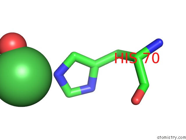
Mono view
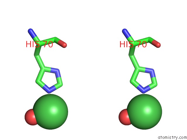
Stereo pair view
Mono view
Stereo pair view
A full contact list of Nickel with other atoms in the Ni binding
site number 1 of Crystal Structure of Spectinomycin Phosphotransferase, Aph(9)-Ia, in Complex with Adp and Spectinomcyin within 5.0Å range:
|
Reference:
D.H.Fong,
C.T.Lemke,
J.Hwang,
B.Xiong,
A.M.Berghuis.
Structure of the Antibiotic Resistance Factor Spectinomycin Phosphotransferase From Legionella Pneumophila. J.Biol.Chem. V. 285 9545 2010.
ISSN: ISSN 0021-9258
PubMed: 20089863
DOI: 10.1074/JBC.M109.038364
Page generated: Wed Oct 9 17:22:57 2024
ISSN: ISSN 0021-9258
PubMed: 20089863
DOI: 10.1074/JBC.M109.038364
Last articles
Zn in 9J0NZn in 9J0O
Zn in 9J0P
Zn in 9FJX
Zn in 9EKB
Zn in 9C0F
Zn in 9CAH
Zn in 9CH0
Zn in 9CH3
Zn in 9CH1