Nickel »
PDB 3hy4-3kvb »
3imp »
Nickel in PDB 3imp: New Crystal Form of the C-Terminal Domain of Helicobacter Pylori Motb (Residues 125-256)
Protein crystallography data
The structure of New Crystal Form of the C-Terminal Domain of Helicobacter Pylori Motb (Residues 125-256), PDB code: 3imp
was solved by
A.Roujeinikova,
with X-Ray Crystallography technique. A brief refinement statistics is given in the table below:
| Resolution Low / High (Å) | 15.00 / 2.50 |
| Space group | P 1 21 1 |
| Cell size a, b, c (Å), α, β, γ (°) | 107.560, 100.336, 108.490, 90.00, 119.51, 90.00 |
| R / Rfree (%) | 18.6 / 24.9 |
Other elements in 3imp:
The structure of New Crystal Form of the C-Terminal Domain of Helicobacter Pylori Motb (Residues 125-256) also contains other interesting chemical elements:
| Chlorine | (Cl) | 4 atoms |
Nickel Binding Sites:
The binding sites of Nickel atom in the New Crystal Form of the C-Terminal Domain of Helicobacter Pylori Motb (Residues 125-256)
(pdb code 3imp). This binding sites where shown within
5.0 Angstroms radius around Nickel atom.
In total 2 binding sites of Nickel where determined in the New Crystal Form of the C-Terminal Domain of Helicobacter Pylori Motb (Residues 125-256), PDB code: 3imp:
Jump to Nickel binding site number: 1; 2;
In total 2 binding sites of Nickel where determined in the New Crystal Form of the C-Terminal Domain of Helicobacter Pylori Motb (Residues 125-256), PDB code: 3imp:
Jump to Nickel binding site number: 1; 2;
Nickel binding site 1 out of 2 in 3imp
Go back to
Nickel binding site 1 out
of 2 in the New Crystal Form of the C-Terminal Domain of Helicobacter Pylori Motb (Residues 125-256)
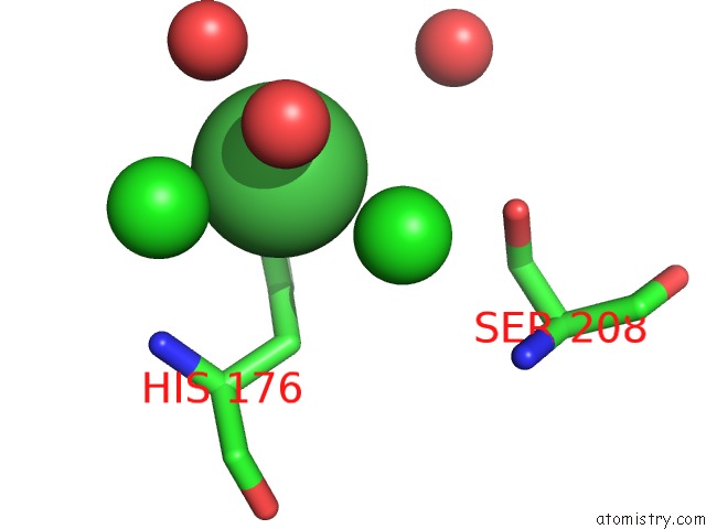
Mono view
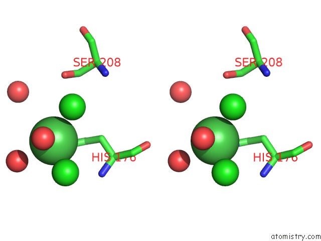
Stereo pair view
Mono view
Stereo pair view
A full contact list of Nickel with other atoms in the Ni binding
site number 1 of New Crystal Form of the C-Terminal Domain of Helicobacter Pylori Motb (Residues 125-256) within 5.0Å range:
|
Nickel binding site 2 out of 2 in 3imp
Go back to
Nickel binding site 2 out
of 2 in the New Crystal Form of the C-Terminal Domain of Helicobacter Pylori Motb (Residues 125-256)
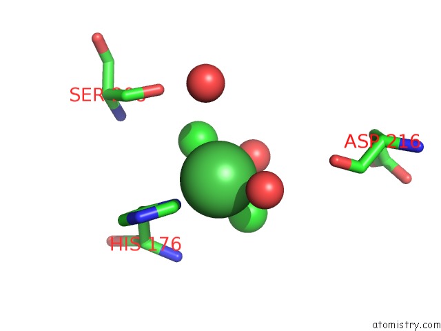
Mono view
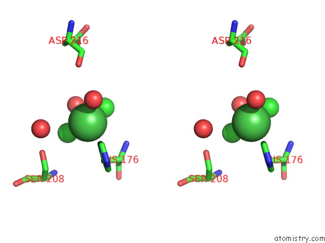
Stereo pair view
Mono view
Stereo pair view
A full contact list of Nickel with other atoms in the Ni binding
site number 2 of New Crystal Form of the C-Terminal Domain of Helicobacter Pylori Motb (Residues 125-256) within 5.0Å range:
|
Reference:
C.F.Reboul,
D.A.Andrews,
M.F.Nahar,
A.M.Buckle,
A.Roujeinikova.
Crystallographic and Molecular Dynamics Analysis of Loop Motions Unmasking the Peptidoglycan-Binding Site in Stator Protein Motb of Flagellar Motor Plos One V. 6 E1898 2011.
ISSN: ESSN 1932-6203
PubMed: 21533052
DOI: 10.1371/JOURNAL.PONE.0018981
Page generated: Wed Oct 9 17:24:35 2024
ISSN: ESSN 1932-6203
PubMed: 21533052
DOI: 10.1371/JOURNAL.PONE.0018981
Last articles
Zn in 9J0NZn in 9J0O
Zn in 9J0P
Zn in 9FJX
Zn in 9EKB
Zn in 9C0F
Zn in 9CAH
Zn in 9CH0
Zn in 9CH3
Zn in 9CH1