Nickel »
PDB 3hy4-3kvb »
3kco »
Nickel in PDB 3kco: Room Temperature Neutron Structure of D-Xylose Isomerase in Complex with Two NI2+ Cations and D12-D-Glucose in the Linear Form (Refined Jointly with X-Ray Structure 3KBN)
Enzymatic activity of Room Temperature Neutron Structure of D-Xylose Isomerase in Complex with Two NI2+ Cations and D12-D-Glucose in the Linear Form (Refined Jointly with X-Ray Structure 3KBN)
All present enzymatic activity of Room Temperature Neutron Structure of D-Xylose Isomerase in Complex with Two NI2+ Cations and D12-D-Glucose in the Linear Form (Refined Jointly with X-Ray Structure 3KBN):
5.3.1.5;
5.3.1.5;
Protein crystallography data
The structure of Room Temperature Neutron Structure of D-Xylose Isomerase in Complex with Two NI2+ Cations and D12-D-Glucose in the Linear Form (Refined Jointly with X-Ray Structure 3KBN), PDB code: 3kco
was solved by
A.Y.Kovalevsky,
P.Langan,
with X-Ray Crystallography technique. A brief refinement statistics is given in the table below:
| Resolution Low / High (Å) | N/A / 1.80 |
| Space group | I 2 2 2 |
| Cell size a, b, c (Å), α, β, γ (°) | 94.007, 99.669, 102.862, 90.00, 90.00, 90.00 |
| R / Rfree (%) | 19.9 / 21.1 |
Nickel Binding Sites:
The binding sites of Nickel atom in the Room Temperature Neutron Structure of D-Xylose Isomerase in Complex with Two NI2+ Cations and D12-D-Glucose in the Linear Form (Refined Jointly with X-Ray Structure 3KBN)
(pdb code 3kco). This binding sites where shown within
5.0 Angstroms radius around Nickel atom.
In total 3 binding sites of Nickel where determined in the Room Temperature Neutron Structure of D-Xylose Isomerase in Complex with Two NI2+ Cations and D12-D-Glucose in the Linear Form (Refined Jointly with X-Ray Structure 3KBN), PDB code: 3kco:
Jump to Nickel binding site number: 1; 2; 3;
In total 3 binding sites of Nickel where determined in the Room Temperature Neutron Structure of D-Xylose Isomerase in Complex with Two NI2+ Cations and D12-D-Glucose in the Linear Form (Refined Jointly with X-Ray Structure 3KBN), PDB code: 3kco:
Jump to Nickel binding site number: 1; 2; 3;
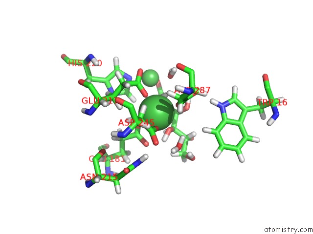
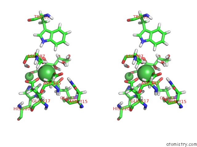
Nickel binding site 1 out of 3 in 3kco
Go back to
Nickel binding site 1 out
of 3 in the Room Temperature Neutron Structure of D-Xylose Isomerase in Complex with Two NI2+ Cations and D12-D-Glucose in the Linear Form (Refined Jointly with X-Ray Structure 3KBN)
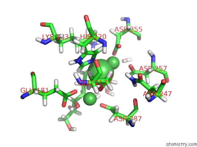
Mono view
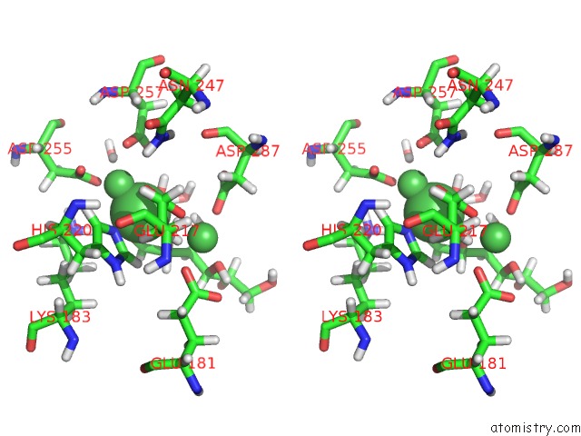
Stereo pair view
Mono view
Stereo pair view
|
|
A full contact list of Nickel with other atoms in the Ni binding
site number 1 of Room Temperature Neutron Structure of D-Xylose Isomerase in Complex with Two NI2+ Cations and D12-D-Glucose in the Linear Form (Refined Jointly with X-Ray Structure 3KBN) within 5.0Å range:
|
Nickel binding site 2 out of 3 in 3kco
Go back to
Nickel binding site 2 out
of 3 in the Room Temperature Neutron Structure of D-Xylose Isomerase in Complex with Two NI2+ Cations and D12-D-Glucose in the Linear Form (Refined Jointly with X-Ray Structure 3KBN)
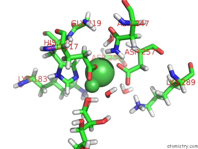
Mono view
Stereo pair view
Mono view
Stereo pair view
|
|
A full contact list of Nickel with other atoms in the Ni binding
site number 2 of Room Temperature Neutron Structure of D-Xylose Isomerase in Complex with Two NI2+ Cations and D12-D-Glucose in the Linear Form (Refined Jointly with X-Ray Structure 3KBN) within 5.0Å range:
|
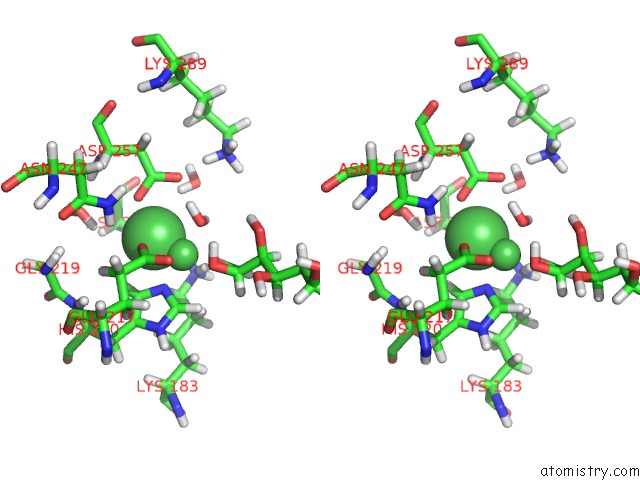
Nickel binding site 3 out of 3 in 3kco
Go back to
Nickel binding site 3 out
of 3 in the Room Temperature Neutron Structure of D-Xylose Isomerase in Complex with Two NI2+ Cations and D12-D-Glucose in the Linear Form (Refined Jointly with X-Ray Structure 3KBN)
Mono view
Stereo pair view
Mono view
Stereo pair view
|
|
A full contact list of Nickel with other atoms in the Ni binding
site number 3 of Room Temperature Neutron Structure of D-Xylose Isomerase in Complex with Two NI2+ Cations and D12-D-Glucose in the Linear Form (Refined Jointly with X-Ray Structure 3KBN) within 5.0Å range:
|
Reference:
A.Y.Kovalevsky,
L.Hanson,
S.Z.Fisher,
M.Mustyakimov,
S.A.Mason,
V.T.Forsyth,
M.P.Blakeley,
D.A.Keen,
T.Wagner,
H.L.Carrell,
A.K.Katz,
J.P.Glusker,
P.Langan.
Metal Ion Roles and the Movement of Hydrogen During Reaction Catalyzed By D-Xylose Isomerase: A Joint X-Ray and Neutron Diffraction Study. Structure V. 18 688 2010.
ISSN: ISSN 0969-2126
PubMed: 20541506
DOI: 10.1016/J.STR.2010.03.011
Page generated: Wed Oct 9 17:27:28 2024
ISSN: ISSN 0969-2126
PubMed: 20541506
DOI: 10.1016/J.STR.2010.03.011
Last articles
Zn in 9MJ5Zn in 9HNW
Zn in 9G0L
Zn in 9FNE
Zn in 9DZN
Zn in 9E0I
Zn in 9D32
Zn in 9DAK
Zn in 8ZXC
Zn in 8ZUF