Nickel »
PDB 3l1m-3n6n »
3le0 »
Nickel in PDB 3le0: Lectin Domain of Lectinolysin Complexed with Glycerol
Protein crystallography data
The structure of Lectin Domain of Lectinolysin Complexed with Glycerol, PDB code: 3le0
was solved by
S.C.Feil,
with X-Ray Crystallography technique. A brief refinement statistics is given in the table below:
| Resolution Low / High (Å) | 29.46 / 1.91 |
| Space group | P 43 21 2 |
| Cell size a, b, c (Å), α, β, γ (°) | 67.189, 67.189, 98.322, 90.00, 90.00, 90.00 |
| R / Rfree (%) | 17.5 / 20.6 |
Other elements in 3le0:
The structure of Lectin Domain of Lectinolysin Complexed with Glycerol also contains other interesting chemical elements:
| Calcium | (Ca) | 1 atom |
Nickel Binding Sites:
The binding sites of Nickel atom in the Lectin Domain of Lectinolysin Complexed with Glycerol
(pdb code 3le0). This binding sites where shown within
5.0 Angstroms radius around Nickel atom.
In total only one binding site of Nickel was determined in the Lectin Domain of Lectinolysin Complexed with Glycerol, PDB code: 3le0:
In total only one binding site of Nickel was determined in the Lectin Domain of Lectinolysin Complexed with Glycerol, PDB code: 3le0:
Nickel binding site 1 out of 1 in 3le0
Go back to
Nickel binding site 1 out
of 1 in the Lectin Domain of Lectinolysin Complexed with Glycerol
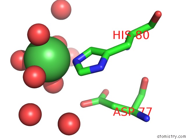
Mono view
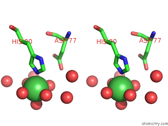
Stereo pair view
Mono view
Stereo pair view
A full contact list of Nickel with other atoms in the Ni binding
site number 1 of Lectin Domain of Lectinolysin Complexed with Glycerol within 5.0Å range:
|
Reference:
S.C.Feil,
S.Lawrence,
T.D.Mulhern,
J.K.Holien,
E.M.Hotze,
S.Farrand,
R.K.Tweten,
M.W.Parker.
Structure of the Lectin Regulatory Domain of the Cholesterol-Dependent Cytolysin Lectinolysin Reveals the Basis For Its Lewis Antigen Specificity. Structure V. 20 248 2012.
ISSN: ISSN 0969-2126
PubMed: 22325774
DOI: 10.1016/J.STR.2011.11.017
Page generated: Wed Oct 9 17:29:41 2024
ISSN: ISSN 0969-2126
PubMed: 22325774
DOI: 10.1016/J.STR.2011.11.017
Last articles
Zn in 9MJ5Zn in 9HNW
Zn in 9G0L
Zn in 9FNE
Zn in 9DZN
Zn in 9E0I
Zn in 9D32
Zn in 9DAK
Zn in 8ZXC
Zn in 8ZUF