Nickel »
PDB 3l1m-3n6n »
3m32 »
Nickel in PDB 3m32: Structural Insight Into Methyl-Coenzyme M Reductase Chemistry Using Coenzyme B Analogues
Enzymatic activity of Structural Insight Into Methyl-Coenzyme M Reductase Chemistry Using Coenzyme B Analogues
All present enzymatic activity of Structural Insight Into Methyl-Coenzyme M Reductase Chemistry Using Coenzyme B Analogues:
2.8.4.1;
2.8.4.1;
Protein crystallography data
The structure of Structural Insight Into Methyl-Coenzyme M Reductase Chemistry Using Coenzyme B Analogues, PDB code: 3m32
was solved by
P.E.Cedervall,
M.Dey,
S.W.Ragsdale,
C.M.Wilmot,
with X-Ray Crystallography technique. A brief refinement statistics is given in the table below:
| Resolution Low / High (Å) | 20.28 / 1.35 |
| Space group | P 1 21 1 |
| Cell size a, b, c (Å), α, β, γ (°) | 82.016, 118.260, 122.387, 90.00, 91.84, 90.00 |
| R / Rfree (%) | 14 / 15.7 |
Other elements in 3m32:
The structure of Structural Insight Into Methyl-Coenzyme M Reductase Chemistry Using Coenzyme B Analogues also contains other interesting chemical elements:
| Magnesium | (Mg) | 9 atoms |
| Zinc | (Zn) | 1 atom |
Nickel Binding Sites:
The binding sites of Nickel atom in the Structural Insight Into Methyl-Coenzyme M Reductase Chemistry Using Coenzyme B Analogues
(pdb code 3m32). This binding sites where shown within
5.0 Angstroms radius around Nickel atom.
In total 2 binding sites of Nickel where determined in the Structural Insight Into Methyl-Coenzyme M Reductase Chemistry Using Coenzyme B Analogues, PDB code: 3m32:
Jump to Nickel binding site number: 1; 2;
In total 2 binding sites of Nickel where determined in the Structural Insight Into Methyl-Coenzyme M Reductase Chemistry Using Coenzyme B Analogues, PDB code: 3m32:
Jump to Nickel binding site number: 1; 2;
Nickel binding site 1 out of 2 in 3m32
Go back to
Nickel binding site 1 out
of 2 in the Structural Insight Into Methyl-Coenzyme M Reductase Chemistry Using Coenzyme B Analogues
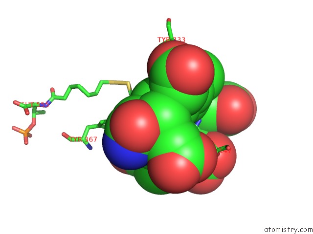
Mono view
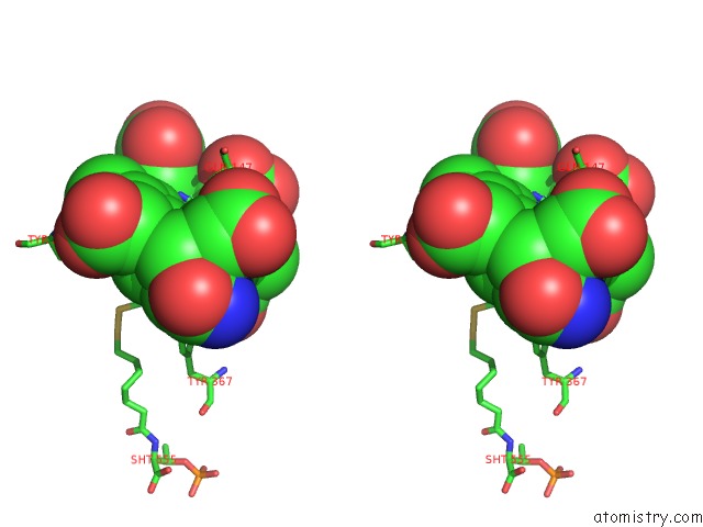
Stereo pair view
Mono view
Stereo pair view
A full contact list of Nickel with other atoms in the Ni binding
site number 1 of Structural Insight Into Methyl-Coenzyme M Reductase Chemistry Using Coenzyme B Analogues within 5.0Å range:
|
Nickel binding site 2 out of 2 in 3m32
Go back to
Nickel binding site 2 out
of 2 in the Structural Insight Into Methyl-Coenzyme M Reductase Chemistry Using Coenzyme B Analogues
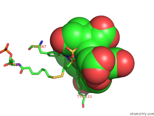
Mono view
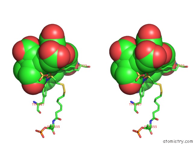
Stereo pair view
Mono view
Stereo pair view
A full contact list of Nickel with other atoms in the Ni binding
site number 2 of Structural Insight Into Methyl-Coenzyme M Reductase Chemistry Using Coenzyme B Analogues within 5.0Å range:
|
Reference:
P.E.Cedervall,
M.Dey,
A.R.Pearson,
S.W.Ragsdale,
C.M.Wilmot.
Structural Insight Into Methyl-Coenzyme M Reductase Chemistry Using Coenzyme B Analogues. Biochemistry V. 49 7683 2010.
ISSN: ISSN 0006-2960
PubMed: 20707311
DOI: 10.1021/BI100458D
Page generated: Wed Oct 9 17:31:15 2024
ISSN: ISSN 0006-2960
PubMed: 20707311
DOI: 10.1021/BI100458D
Last articles
Zn in 9MJ5Zn in 9HNW
Zn in 9G0L
Zn in 9FNE
Zn in 9DZN
Zn in 9E0I
Zn in 9D32
Zn in 9DAK
Zn in 8ZXC
Zn in 8ZUF