Nickel »
PDB 3l1m-3n6n »
3ms5 »
Nickel in PDB 3ms5: Crystal Structure of Human Gamma-Butyrobetaine,2-Oxoglutarate Dioxygenase 1 (BBOX1)
Enzymatic activity of Crystal Structure of Human Gamma-Butyrobetaine,2-Oxoglutarate Dioxygenase 1 (BBOX1)
All present enzymatic activity of Crystal Structure of Human Gamma-Butyrobetaine,2-Oxoglutarate Dioxygenase 1 (BBOX1):
1.14.11.1;
1.14.11.1;
Protein crystallography data
The structure of Crystal Structure of Human Gamma-Butyrobetaine,2-Oxoglutarate Dioxygenase 1 (BBOX1), PDB code: 3ms5
was solved by
T.Krojer,
G.Kochan,
M.A.Mcdonough,
F.Von Delft,
I.K.H.Leung,
L.Henry,
T.D.W.Claridge,
E.Pilka,
E.Ugochukwu,
J.Muniz,
P.Filippakopoulos,
C.Bountra,
C.H.Arrowsmith,
J.Weigelt,
A.Edwards,
K.L.Kavanagh,
C.J.Schofield,
U.Oppermann,
Structural Genomics Consortium (Sgc),
with X-Ray Crystallography technique. A brief refinement statistics is given in the table below:
| Resolution Low / High (Å) | 19.45 / 1.82 |
| Space group | H 3 2 |
| Cell size a, b, c (Å), α, β, γ (°) | 107.366, 107.366, 205.054, 90.00, 90.00, 120.00 |
| R / Rfree (%) | 14.7 / 18.3 |
Other elements in 3ms5:
The structure of Crystal Structure of Human Gamma-Butyrobetaine,2-Oxoglutarate Dioxygenase 1 (BBOX1) also contains other interesting chemical elements:
| Zinc | (Zn) | 1 atom |
Nickel Binding Sites:
The binding sites of Nickel atom in the Crystal Structure of Human Gamma-Butyrobetaine,2-Oxoglutarate Dioxygenase 1 (BBOX1)
(pdb code 3ms5). This binding sites where shown within
5.0 Angstroms radius around Nickel atom.
In total only one binding site of Nickel was determined in the Crystal Structure of Human Gamma-Butyrobetaine,2-Oxoglutarate Dioxygenase 1 (BBOX1), PDB code: 3ms5:
In total only one binding site of Nickel was determined in the Crystal Structure of Human Gamma-Butyrobetaine,2-Oxoglutarate Dioxygenase 1 (BBOX1), PDB code: 3ms5:
Nickel binding site 1 out of 1 in 3ms5
Go back to
Nickel binding site 1 out
of 1 in the Crystal Structure of Human Gamma-Butyrobetaine,2-Oxoglutarate Dioxygenase 1 (BBOX1)
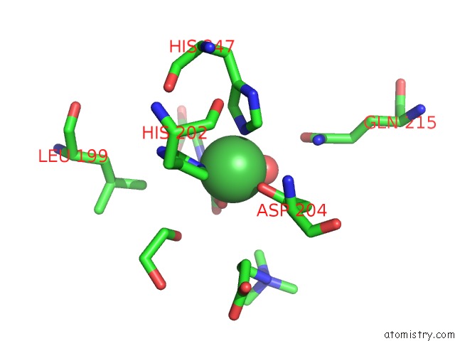
Mono view
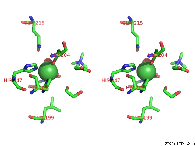
Stereo pair view
Mono view
Stereo pair view
A full contact list of Nickel with other atoms in the Ni binding
site number 1 of Crystal Structure of Human Gamma-Butyrobetaine,2-Oxoglutarate Dioxygenase 1 (BBOX1) within 5.0Å range:
|
Reference:
I.K.Leung,
T.J.Krojer,
G.T.Kochan,
L.Henry,
F.Von Delft,
T.D.Claridge,
U.Oppermann,
M.A.Mcdonough,
C.J.Schofield.
Structural and Mechanistic Studies on Gamma-Butyrobetaine Hydroxylase. Chem. Biol. V. 17 1316 2010.
ISSN: ISSN 1879-1301
PubMed: 21168767
DOI: 10.1016/J.CHEMBIOL.2010.09.016
Page generated: Wed Oct 9 17:32:41 2024
ISSN: ISSN 1879-1301
PubMed: 21168767
DOI: 10.1016/J.CHEMBIOL.2010.09.016
Last articles
Zn in 9MJ5Zn in 9HNW
Zn in 9G0L
Zn in 9FNE
Zn in 9DZN
Zn in 9E0I
Zn in 9D32
Zn in 9DAK
Zn in 8ZXC
Zn in 8ZUF