Nickel »
PDB 3qsi-3tsn »
3s0k »
Nickel in PDB 3s0k: Crystal Structure of Human Glycolipid Transfer Protein Complexed with Glucosylceramide Containing Oleoyl Acyl Chain (18:1)
Protein crystallography data
The structure of Crystal Structure of Human Glycolipid Transfer Protein Complexed with Glucosylceramide Containing Oleoyl Acyl Chain (18:1), PDB code: 3s0k
was solved by
A.Cabo-Bilbao,
V.Samygina,
A.N.Popov,
B.Ochoa-Lizarralde,
F.Goni-De-Cerio,
D.J.Patel,
R.E.Brown,
L.Malinina,
with X-Ray Crystallography technique. A brief refinement statistics is given in the table below:
| Resolution Low / High (Å) | 15.00 / 1.40 |
| Space group | P 21 21 21 |
| Cell size a, b, c (Å), α, β, γ (°) | 50.659, 61.646, 67.214, 90.00, 90.00, 90.00 |
| R / Rfree (%) | 15 / 18.2 |
Nickel Binding Sites:
The binding sites of Nickel atom in the Crystal Structure of Human Glycolipid Transfer Protein Complexed with Glucosylceramide Containing Oleoyl Acyl Chain (18:1)
(pdb code 3s0k). This binding sites where shown within
5.0 Angstroms radius around Nickel atom.
In total only one binding site of Nickel was determined in the Crystal Structure of Human Glycolipid Transfer Protein Complexed with Glucosylceramide Containing Oleoyl Acyl Chain (18:1), PDB code: 3s0k:
In total only one binding site of Nickel was determined in the Crystal Structure of Human Glycolipid Transfer Protein Complexed with Glucosylceramide Containing Oleoyl Acyl Chain (18:1), PDB code: 3s0k:
Nickel binding site 1 out of 1 in 3s0k
Go back to
Nickel binding site 1 out
of 1 in the Crystal Structure of Human Glycolipid Transfer Protein Complexed with Glucosylceramide Containing Oleoyl Acyl Chain (18:1)
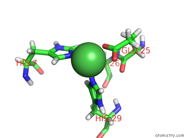
Mono view
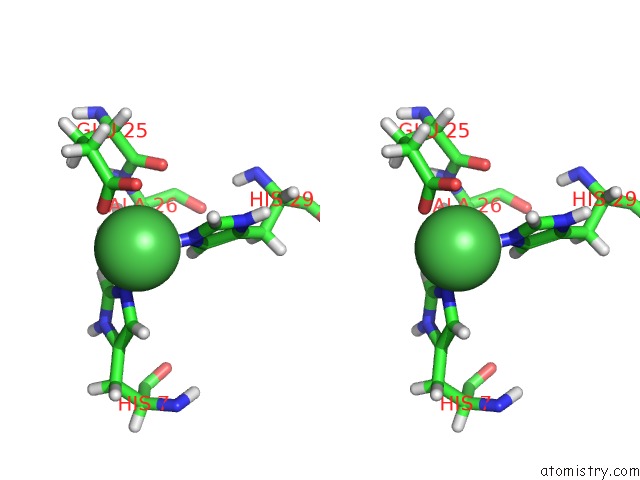
Stereo pair view
Mono view
Stereo pair view
A full contact list of Nickel with other atoms in the Ni binding
site number 1 of Crystal Structure of Human Glycolipid Transfer Protein Complexed with Glucosylceramide Containing Oleoyl Acyl Chain (18:1) within 5.0Å range:
|
Reference:
V.R.Samygina,
A.N.Popov,
A.Cabo-Bilbao,
B.Ochoa-Lizarralde,
F.Goni-De-Cerio,
X.Zhai,
J.G.Molotkovsky,
D.J.Patel,
R.E.Brown,
L.Malinina.
Enhanced Selectivity For Sulfatide By Engineered Human Glycolipid Transfer Protein. Structure V. 19 1644 2011.
ISSN: ISSN 0969-2126
PubMed: 22078563
DOI: 10.1016/J.STR.2011.09.011
Page generated: Wed Oct 9 17:46:52 2024
ISSN: ISSN 0969-2126
PubMed: 22078563
DOI: 10.1016/J.STR.2011.09.011
Last articles
Zn in 9J0NZn in 9J0O
Zn in 9J0P
Zn in 9FJX
Zn in 9EKB
Zn in 9C0F
Zn in 9CAH
Zn in 9CH0
Zn in 9CH3
Zn in 9CH1