Nickel »
PDB 3qsi-3tsn »
3s2x »
Nickel in PDB 3s2x: Structure of Acetyl-Coenzyme A Synthase Alpha Subunit C-Terminal Domain
Enzymatic activity of Structure of Acetyl-Coenzyme A Synthase Alpha Subunit C-Terminal Domain
All present enzymatic activity of Structure of Acetyl-Coenzyme A Synthase Alpha Subunit C-Terminal Domain:
2.3.1.169;
2.3.1.169;
Protein crystallography data
The structure of Structure of Acetyl-Coenzyme A Synthase Alpha Subunit C-Terminal Domain, PDB code: 3s2x
was solved by
P.Li,
with X-Ray Crystallography technique. A brief refinement statistics is given in the table below:
| Resolution Low / High (Å) | 42.60 / 2.35 |
| Space group | H 3 |
| Cell size a, b, c (Å), α, β, γ (°) | 129.040, 129.040, 46.030, 90.00, 90.00, 120.00 |
| R / Rfree (%) | 22.1 / 26.4 |
Nickel Binding Sites:
The binding sites of Nickel atom in the Structure of Acetyl-Coenzyme A Synthase Alpha Subunit C-Terminal Domain
(pdb code 3s2x). This binding sites where shown within
5.0 Angstroms radius around Nickel atom.
In total 4 binding sites of Nickel where determined in the Structure of Acetyl-Coenzyme A Synthase Alpha Subunit C-Terminal Domain, PDB code: 3s2x:
Jump to Nickel binding site number: 1; 2; 3; 4;
In total 4 binding sites of Nickel where determined in the Structure of Acetyl-Coenzyme A Synthase Alpha Subunit C-Terminal Domain, PDB code: 3s2x:
Jump to Nickel binding site number: 1; 2; 3; 4;
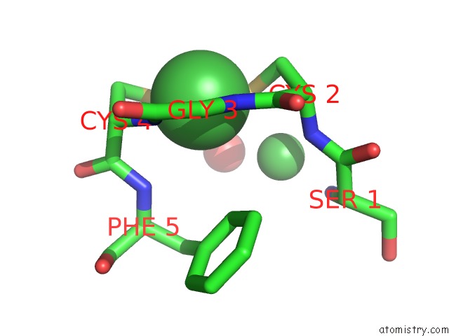
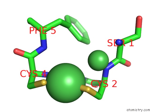
Nickel binding site 1 out of 4 in 3s2x
Go back to
Nickel binding site 1 out
of 4 in the Structure of Acetyl-Coenzyme A Synthase Alpha Subunit C-Terminal Domain
Mono view
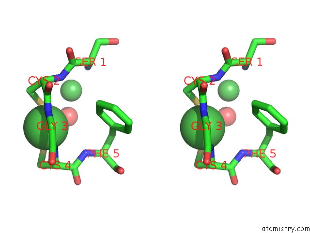
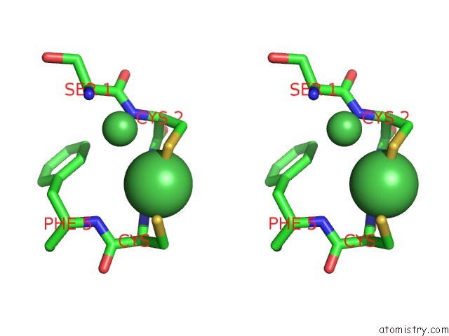
Stereo pair view
Mono view
Stereo pair view
A full contact list of Nickel with other atoms in the Ni binding
site number 1 of Structure of Acetyl-Coenzyme A Synthase Alpha Subunit C-Terminal Domain within 5.0Å range:
|
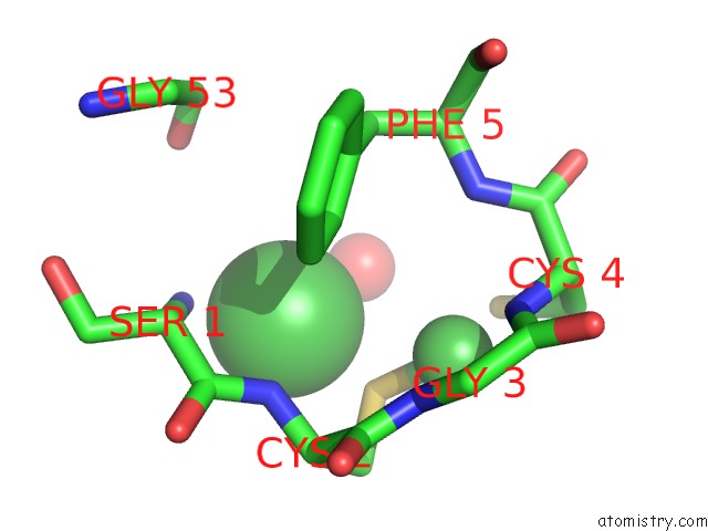
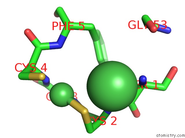
Nickel binding site 2 out of 4 in 3s2x
Go back to
Nickel binding site 2 out
of 4 in the Structure of Acetyl-Coenzyme A Synthase Alpha Subunit C-Terminal Domain
Mono view
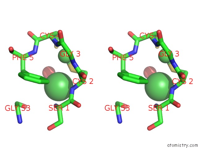
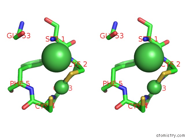
Stereo pair view
Mono view
Stereo pair view
A full contact list of Nickel with other atoms in the Ni binding
site number 2 of Structure of Acetyl-Coenzyme A Synthase Alpha Subunit C-Terminal Domain within 5.0Å range:
|
Nickel binding site 3 out of 4 in 3s2x
Go back to
Nickel binding site 3 out
of 4 in the Structure of Acetyl-Coenzyme A Synthase Alpha Subunit C-Terminal Domain
Mono view
Stereo pair view
Mono view
Stereo pair view
A full contact list of Nickel with other atoms in the Ni binding
site number 3 of Structure of Acetyl-Coenzyme A Synthase Alpha Subunit C-Terminal Domain within 5.0Å range:
|
Nickel binding site 4 out of 4 in 3s2x
Go back to
Nickel binding site 4 out
of 4 in the Structure of Acetyl-Coenzyme A Synthase Alpha Subunit C-Terminal Domain
Mono view
Stereo pair view
Mono view
Stereo pair view
A full contact list of Nickel with other atoms in the Ni binding
site number 4 of Structure of Acetyl-Coenzyme A Synthase Alpha Subunit C-Terminal Domain within 5.0Å range:
|
Reference:
Y.Liu,
F.Wang,
P.Li,
X.Tan.
Insights Into the Mechanistic Role of the [Fe(4) S(4) ] Cubane in the A-Cluster {[Fe(4) S(4) ]-(Sr)-[Ni(P) Ni(D) ]} of Acetyl-Coenzyme A Synthase. Chembiochem V. 12 1417 2011.
ISSN: ISSN 1439-4227
PubMed: 21626638
DOI: 10.1002/CBIC.201100101
Page generated: Mon Aug 18 18:57:43 2025
ISSN: ISSN 1439-4227
PubMed: 21626638
DOI: 10.1002/CBIC.201100101
Last articles
Ni in 5U9ENi in 5TVS
Ni in 5U93
Ni in 5TVR
Ni in 5TRQ
Ni in 5TFZ
Ni in 5TR8
Ni in 5T22
Ni in 5TCW
Ni in 5TBX