Nickel »
PDB 3u4s-446d »
3wrq »
Nickel in PDB 3wrq: Minute Virus of Mice Non-Structural Protein-1N-Terminal Nuclease Domain Reveals A Unique ZN2+ Coordination in the Active Site Pocket and Shows A Novel Mode of Dna Recognition at the Origin of Replication
Protein crystallography data
The structure of Minute Virus of Mice Non-Structural Protein-1N-Terminal Nuclease Domain Reveals A Unique ZN2+ Coordination in the Active Site Pocket and Shows A Novel Mode of Dna Recognition at the Origin of Replication, PDB code: 3wrq
was solved by
S.K.Tewary,
H.Zhao,
L.Tang,
with X-Ray Crystallography technique. A brief refinement statistics is given in the table below:
| Resolution Low / High (Å) | 30.32 / 1.53 |
| Space group | P 21 21 2 |
| Cell size a, b, c (Å), α, β, γ (°) | 57.280, 121.260, 41.650, 90.00, 90.00, 90.00 |
| R / Rfree (%) | 17.4 / 19.9 |
Nickel Binding Sites:
The binding sites of Nickel atom in the Minute Virus of Mice Non-Structural Protein-1N-Terminal Nuclease Domain Reveals A Unique ZN2+ Coordination in the Active Site Pocket and Shows A Novel Mode of Dna Recognition at the Origin of Replication
(pdb code 3wrq). This binding sites where shown within
5.0 Angstroms radius around Nickel atom.
In total only one binding site of Nickel was determined in the Minute Virus of Mice Non-Structural Protein-1N-Terminal Nuclease Domain Reveals A Unique ZN2+ Coordination in the Active Site Pocket and Shows A Novel Mode of Dna Recognition at the Origin of Replication, PDB code: 3wrq:
In total only one binding site of Nickel was determined in the Minute Virus of Mice Non-Structural Protein-1N-Terminal Nuclease Domain Reveals A Unique ZN2+ Coordination in the Active Site Pocket and Shows A Novel Mode of Dna Recognition at the Origin of Replication, PDB code: 3wrq:
Nickel binding site 1 out of 1 in 3wrq
Go back to
Nickel binding site 1 out
of 1 in the Minute Virus of Mice Non-Structural Protein-1N-Terminal Nuclease Domain Reveals A Unique ZN2+ Coordination in the Active Site Pocket and Shows A Novel Mode of Dna Recognition at the Origin of Replication
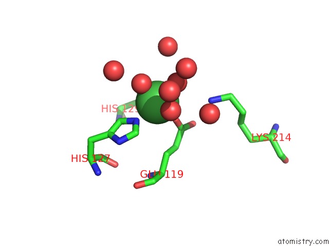
Mono view
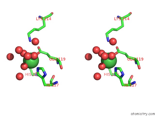
Stereo pair view
Mono view
Stereo pair view
A full contact list of Nickel with other atoms in the Ni binding
site number 1 of Minute Virus of Mice Non-Structural Protein-1N-Terminal Nuclease Domain Reveals A Unique ZN2+ Coordination in the Active Site Pocket and Shows A Novel Mode of Dna Recognition at the Origin of Replication within 5.0Å range:
|
Reference:
S.K.Tewary,
L.Liang,
Z.Lin,
A.Lynn,
S.F.Cotmore,
P.Tattersall,
H.Zhao,
L.Tang.
Structures of Minute Virus of Mice Replication Initiator Protein N-Terminal Domain: Insights Into Dna Nicking and Origin Binding. Virology V.476C 61 2014.
ISSN: ISSN 0042-6822
PubMed: 25528417
DOI: 10.1016/J.VIROL.2014.11.022
Page generated: Wed Oct 9 17:54:24 2024
ISSN: ISSN 0042-6822
PubMed: 25528417
DOI: 10.1016/J.VIROL.2014.11.022
Last articles
Zn in 9J0NZn in 9J0O
Zn in 9J0P
Zn in 9FJX
Zn in 9EKB
Zn in 9C0F
Zn in 9CAH
Zn in 9CH0
Zn in 9CH3
Zn in 9CH1