Nickel »
PDB 4jzr-4ls3 »
4k0o »
Nickel in PDB 4k0o: F17B-G Lectin Domain with Bound Glcnac(BETA1-3)Gal
Protein crystallography data
The structure of F17B-G Lectin Domain with Bound Glcnac(BETA1-3)Gal, PDB code: 4k0o
was solved by
L.Buts,
R.Loris,
J.Bouckaert,
K.Moonens,
with X-Ray Crystallography technique. A brief refinement statistics is given in the table below:
| Resolution Low / High (Å) | 43.82 / 2.15 |
| Space group | P 62 |
| Cell size a, b, c (Å), α, β, γ (°) | 87.646, 87.646, 57.022, 90.00, 90.00, 120.00 |
| R / Rfree (%) | 16.8 / 18 |
Nickel Binding Sites:
The binding sites of Nickel atom in the F17B-G Lectin Domain with Bound Glcnac(BETA1-3)Gal
(pdb code 4k0o). This binding sites where shown within
5.0 Angstroms radius around Nickel atom.
In total 2 binding sites of Nickel where determined in the F17B-G Lectin Domain with Bound Glcnac(BETA1-3)Gal, PDB code: 4k0o:
Jump to Nickel binding site number: 1; 2;
In total 2 binding sites of Nickel where determined in the F17B-G Lectin Domain with Bound Glcnac(BETA1-3)Gal, PDB code: 4k0o:
Jump to Nickel binding site number: 1; 2;
Nickel binding site 1 out of 2 in 4k0o
Go back to
Nickel binding site 1 out
of 2 in the F17B-G Lectin Domain with Bound Glcnac(BETA1-3)Gal
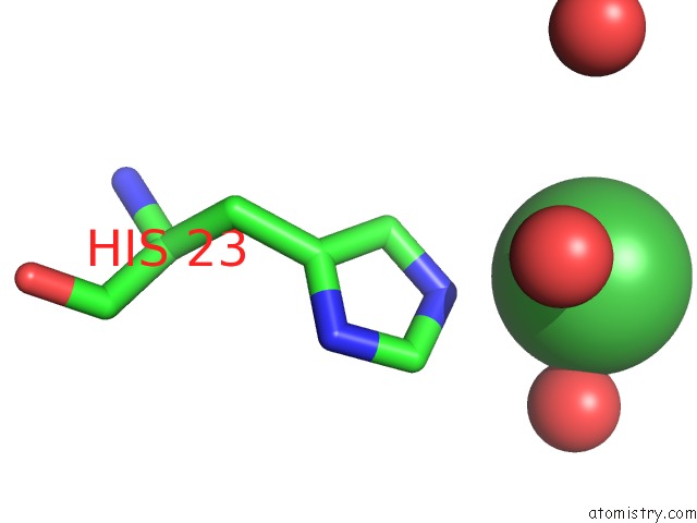
Mono view
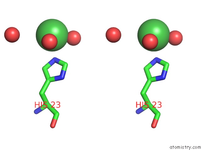
Stereo pair view
Mono view
Stereo pair view
A full contact list of Nickel with other atoms in the Ni binding
site number 1 of F17B-G Lectin Domain with Bound Glcnac(BETA1-3)Gal within 5.0Å range:
|
Nickel binding site 2 out of 2 in 4k0o
Go back to
Nickel binding site 2 out
of 2 in the F17B-G Lectin Domain with Bound Glcnac(BETA1-3)Gal
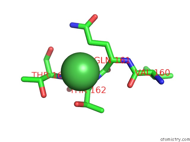
Mono view
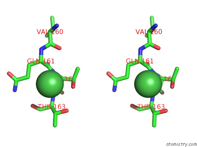
Stereo pair view
Mono view
Stereo pair view
A full contact list of Nickel with other atoms in the Ni binding
site number 2 of F17B-G Lectin Domain with Bound Glcnac(BETA1-3)Gal within 5.0Å range:
|
Reference:
E.Lonardi,
K.Moonens,
L.Buts,
A.R.De Boer,
J.D.Olsson,
M.S.Weiss,
E.Fabre,
Y.Guerardel,
A.M.Deelder,
S.Oscarson,
M.Wuhrer,
J.Bouckaert.
Structural Sampling of Glycan Interaction Profiles Reveals Mucosal Receptors For Fimbrial Adhesins of Enterotoxigenic Escherichia Coli Biology (Basel) V. 2 894 2013.
ISSN: ESSN 2079-7737
PubMed: 24833052
DOI: 10.3390/BIOLOGY2030894
Page generated: Wed Oct 9 18:18:30 2024
ISSN: ESSN 2079-7737
PubMed: 24833052
DOI: 10.3390/BIOLOGY2030894
Last articles
Zn in 9MJ5Zn in 9HNW
Zn in 9G0L
Zn in 9FNE
Zn in 9DZN
Zn in 9E0I
Zn in 9D32
Zn in 9DAK
Zn in 8ZXC
Zn in 8ZUF