Nickel »
PDB 5mdk-5ph1 »
5o5d »
Nickel in PDB 5o5d: Cellobiohydrolase CEL7A From T. Atroviride
Protein crystallography data
The structure of Cellobiohydrolase CEL7A From T. Atroviride, PDB code: 5o5d
was solved by
A.S.Borisova,
J.Stahlberg,
H.Hansson,
with X-Ray Crystallography technique. A brief refinement statistics is given in the table below:
| Resolution Low / High (Å) | 34.74 / 1.72 |
| Space group | P 1 21 1 |
| Cell size a, b, c (Å), α, β, γ (°) | 56.170, 71.670, 104.220, 90.00, 90.00, 90.00 |
| R / Rfree (%) | 14.8 / 17.3 |
Other elements in 5o5d:
The structure of Cellobiohydrolase CEL7A From T. Atroviride also contains other interesting chemical elements:
| Chlorine | (Cl) | 4 atoms |
Nickel Binding Sites:
The binding sites of Nickel atom in the Cellobiohydrolase CEL7A From T. Atroviride
(pdb code 5o5d). This binding sites where shown within
5.0 Angstroms radius around Nickel atom.
In total 4 binding sites of Nickel where determined in the Cellobiohydrolase CEL7A From T. Atroviride, PDB code: 5o5d:
Jump to Nickel binding site number: 1; 2; 3; 4;
In total 4 binding sites of Nickel where determined in the Cellobiohydrolase CEL7A From T. Atroviride, PDB code: 5o5d:
Jump to Nickel binding site number: 1; 2; 3; 4;
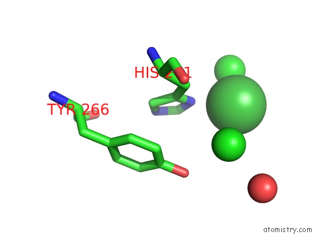
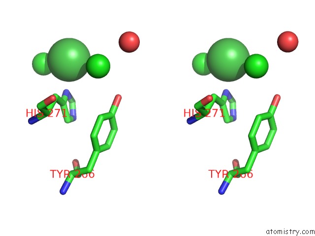
Nickel binding site 1 out of 4 in 5o5d
Go back to
Nickel binding site 1 out
of 4 in the Cellobiohydrolase CEL7A From T. Atroviride
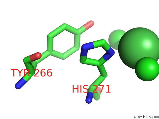
Mono view
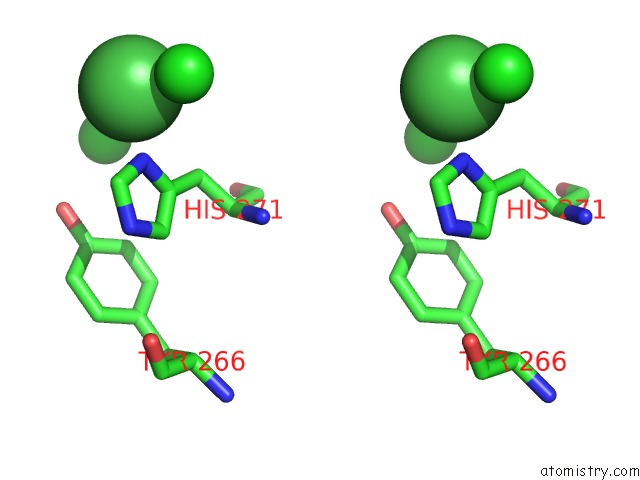
Stereo pair view
Mono view
Stereo pair view
A full contact list of Nickel with other atoms in the Ni binding
site number 1 of Cellobiohydrolase CEL7A From T. Atroviride within 5.0Å range:
|
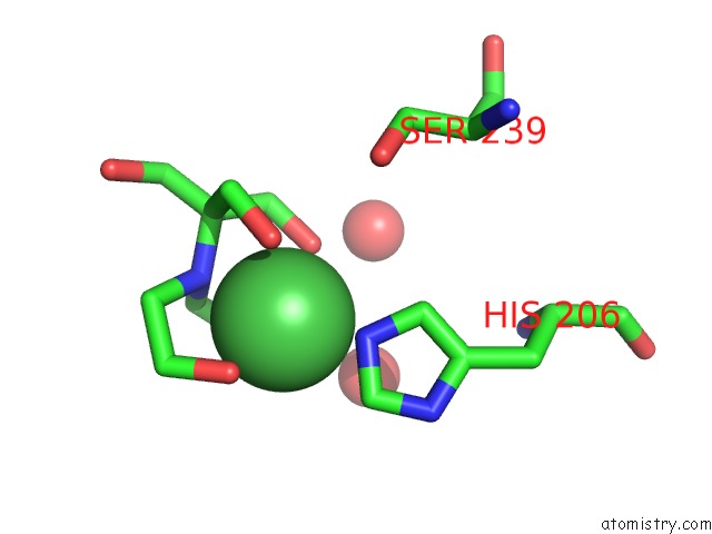
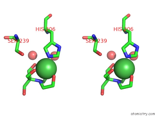
Nickel binding site 2 out of 4 in 5o5d
Go back to
Nickel binding site 2 out
of 4 in the Cellobiohydrolase CEL7A From T. Atroviride
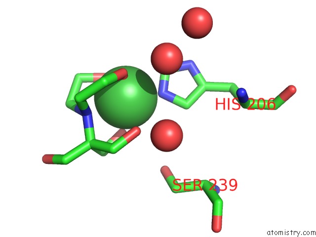
Mono view
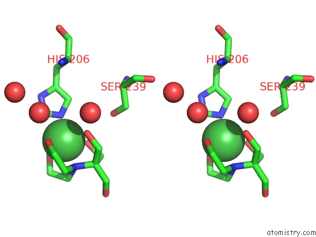
Stereo pair view
Mono view
Stereo pair view
A full contact list of Nickel with other atoms in the Ni binding
site number 2 of Cellobiohydrolase CEL7A From T. Atroviride within 5.0Å range:
|
Nickel binding site 3 out of 4 in 5o5d
Go back to
Nickel binding site 3 out
of 4 in the Cellobiohydrolase CEL7A From T. Atroviride
Mono view
Stereo pair view
Mono view
Stereo pair view
A full contact list of Nickel with other atoms in the Ni binding
site number 3 of Cellobiohydrolase CEL7A From T. Atroviride within 5.0Å range:
|
Nickel binding site 4 out of 4 in 5o5d
Go back to
Nickel binding site 4 out
of 4 in the Cellobiohydrolase CEL7A From T. Atroviride
Mono view
Stereo pair view
Mono view
Stereo pair view
A full contact list of Nickel with other atoms in the Ni binding
site number 4 of Cellobiohydrolase CEL7A From T. Atroviride within 5.0Å range:
|
Reference:
A.S.Borisova,
E.V.Eneyskaya,
S.Jana,
S.F.Badino,
J.Kari,
A.Amore,
M.Karlsson,
H.Hansson,
M.Sandgren,
M.E.Himmel,
P.Westh,
C.M.Payne,
A.A.Kulminskaya,
J.Stahlberg.
Correlation of Structure, Function and Protein Dynamics in GH7 Cellobiohydrolases From Trichoderma Atroviride, T. Reesei and T. Harzianum. Biotechnol Biofuels V. 11 5 2018.
ISSN: ESSN 1754-6834
PubMed: 29344086
DOI: 10.1186/S13068-017-1006-7
Page generated: Thu Oct 10 06:42:13 2024
ISSN: ESSN 1754-6834
PubMed: 29344086
DOI: 10.1186/S13068-017-1006-7
Last articles
Zn in 9J0NZn in 9J0O
Zn in 9J0P
Zn in 9FJX
Zn in 9EKB
Zn in 9C0F
Zn in 9CAH
Zn in 9CH0
Zn in 9CH3
Zn in 9CH1