Nickel »
PDB 5mdk-5ph1 »
5o9w »
Nickel in PDB 5o9w: Thebaine 6-O-Demethylase (T6ODM) From Papaver Somniferum in Complex with 2-Oxoglutarate
Enzymatic activity of Thebaine 6-O-Demethylase (T6ODM) From Papaver Somniferum in Complex with 2-Oxoglutarate
All present enzymatic activity of Thebaine 6-O-Demethylase (T6ODM) From Papaver Somniferum in Complex with 2-Oxoglutarate:
1.14.11.31;
1.14.11.31;
Protein crystallography data
The structure of Thebaine 6-O-Demethylase (T6ODM) From Papaver Somniferum in Complex with 2-Oxoglutarate, PDB code: 5o9w
was solved by
A.Kluza,
E.Niedzialkowska,
K.Kurpiewska,
P.J.Porebski,
T.Borowski,
with X-Ray Crystallography technique. A brief refinement statistics is given in the table below:
| Resolution Low / High (Å) | 41.43 / 1.85 |
| Space group | P 21 21 21 |
| Cell size a, b, c (Å), α, β, γ (°) | 47.044, 87.424, 94.863, 90.00, 90.00, 90.00 |
| R / Rfree (%) | 16.2 / 21.4 |
Other elements in 5o9w:
The structure of Thebaine 6-O-Demethylase (T6ODM) From Papaver Somniferum in Complex with 2-Oxoglutarate also contains other interesting chemical elements:
| Sodium | (Na) | 1 atom |
Nickel Binding Sites:
The binding sites of Nickel atom in the Thebaine 6-O-Demethylase (T6ODM) From Papaver Somniferum in Complex with 2-Oxoglutarate
(pdb code 5o9w). This binding sites where shown within
5.0 Angstroms radius around Nickel atom.
In total only one binding site of Nickel was determined in the Thebaine 6-O-Demethylase (T6ODM) From Papaver Somniferum in Complex with 2-Oxoglutarate, PDB code: 5o9w:
In total only one binding site of Nickel was determined in the Thebaine 6-O-Demethylase (T6ODM) From Papaver Somniferum in Complex with 2-Oxoglutarate, PDB code: 5o9w:
Nickel binding site 1 out of 1 in 5o9w
Go back to
Nickel binding site 1 out
of 1 in the Thebaine 6-O-Demethylase (T6ODM) From Papaver Somniferum in Complex with 2-Oxoglutarate
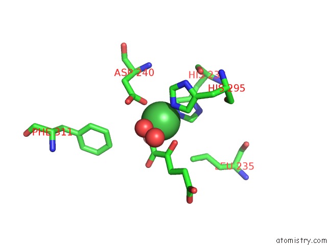
Mono view
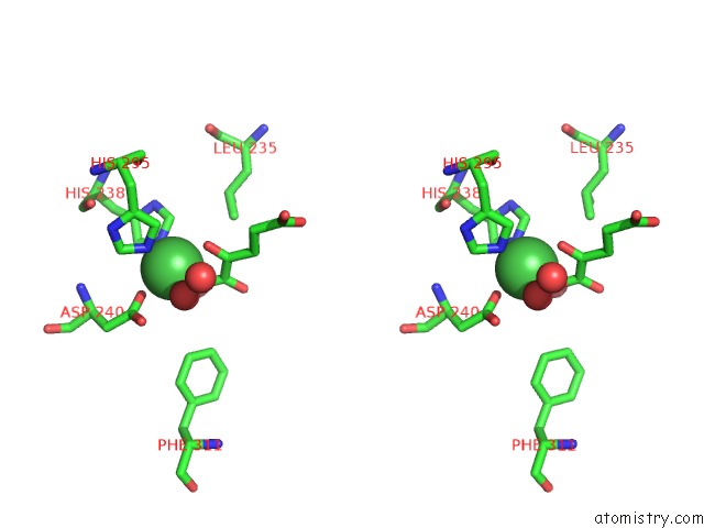
Stereo pair view
Mono view
Stereo pair view
A full contact list of Nickel with other atoms in the Ni binding
site number 1 of Thebaine 6-O-Demethylase (T6ODM) From Papaver Somniferum in Complex with 2-Oxoglutarate within 5.0Å range:
|
Reference:
A.Kluza,
E.Niedzialkowska,
K.Kurpiewska,
Z.Wojdyla,
M.Quesne,
E.Kot,
P.J.Porebski,
T.Borowski.
Crystal Structure of Thebaine 6-O-Demethylase From the Morphine Biosynthesis Pathway. J. Struct. Biol. V. 202 229 2018.
ISSN: ESSN 1095-8657
PubMed: 29408320
DOI: 10.1016/J.JSB.2018.01.007
Page generated: Thu Oct 10 06:42:38 2024
ISSN: ESSN 1095-8657
PubMed: 29408320
DOI: 10.1016/J.JSB.2018.01.007
Last articles
Zn in 9J0NZn in 9J0O
Zn in 9J0P
Zn in 9FJX
Zn in 9EKB
Zn in 9C0F
Zn in 9CAH
Zn in 9CH0
Zn in 9CH3
Zn in 9CH1