Nickel in PDB 5x2u: Direct Observation of Conformational Population Shifts in Hemoglobin: Crystal Structure of Half-Liganded Hemoglobin After Adding 80 Mm Phosphate pH 6.7.
Protein crystallography data
The structure of Direct Observation of Conformational Population Shifts in Hemoglobin: Crystal Structure of Half-Liganded Hemoglobin After Adding 80 Mm Phosphate pH 6.7., PDB code: 5x2u
was solved by
M.Ohki,
S.-Y.Park,
with X-Ray Crystallography technique. A brief refinement statistics is given in the table below:
Resolution Low / High (Å)
|
39.74 /
2.53
|
Space group
|
C 1 2 1
|
Cell size a, b, c (Å), α, β, γ (°)
|
228.886,
55.812,
139.963,
90.00,
102.39,
90.00
|
R / Rfree (%)
|
23.1 /
27.4
|
Other elements in 5x2u:
The structure of Direct Observation of Conformational Population Shifts in Hemoglobin: Crystal Structure of Half-Liganded Hemoglobin After Adding 80 Mm Phosphate pH 6.7. also contains other interesting chemical elements:
Nickel Binding Sites:
The binding sites of Nickel atom in the Direct Observation of Conformational Population Shifts in Hemoglobin: Crystal Structure of Half-Liganded Hemoglobin After Adding 80 Mm Phosphate pH 6.7.
(pdb code 5x2u). This binding sites where shown within
5.0 Angstroms radius around Nickel atom.
In total 6 binding sites of Nickel where determined in the
Direct Observation of Conformational Population Shifts in Hemoglobin: Crystal Structure of Half-Liganded Hemoglobin After Adding 80 Mm Phosphate pH 6.7., PDB code: 5x2u:
Jump to Nickel binding site number:
1;
2;
3;
4;
5;
6;
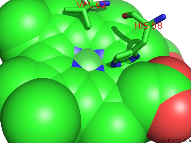
Nickel binding site 1 out
of 6 in 5x2u
Go back to
Nickel Binding Sites List in 5x2u
Nickel binding site 1 out
of 6 in the Direct Observation of Conformational Population Shifts in Hemoglobin: Crystal Structure of Half-Liganded Hemoglobin After Adding 80 Mm Phosphate pH 6.7.
Mono view
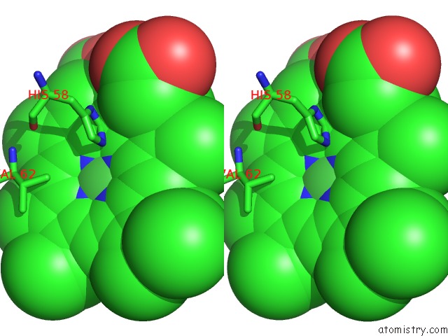
Stereo pair view
|
A full contact list of Nickel with other atoms in the Ni binding
site number 1 of Direct Observation of Conformational Population Shifts in Hemoglobin: Crystal Structure of Half-Liganded Hemoglobin After Adding 80 Mm Phosphate pH 6.7. within 5.0Å range:
probe
|
atom
|
residue
|
distance (Å)
|
B
|
Occ
|
A:Ni201
b:59.5
occ:1.00
|
NI
|
A:HNI201
|
0.0
|
59.5
|
1.0
|
ND
|
A:HNI201
|
1.9
|
60.5
|
1.0
|
NA
|
A:HNI201
|
2.0
|
59.9
|
1.0
|
NB
|
A:HNI201
|
2.0
|
58.5
|
1.0
|
NC
|
A:HNI201
|
2.0
|
59.2
|
1.0
|
NE2
|
A:HIS87
|
2.2
|
83.5
|
1.0
|
C1D
|
A:HNI201
|
3.0
|
61.2
|
1.0
|
C4D
|
A:HNI201
|
3.0
|
61.3
|
1.0
|
C4B
|
A:HNI201
|
3.0
|
58.4
|
1.0
|
C1B
|
A:HNI201
|
3.0
|
58.8
|
1.0
|
C1C
|
A:HNI201
|
3.0
|
58.6
|
1.0
|
C4A
|
A:HNI201
|
3.0
|
59.6
|
1.0
|
C4C
|
A:HNI201
|
3.0
|
59.9
|
1.0
|
C1A
|
A:HNI201
|
3.0
|
60.5
|
1.0
|
CD2
|
A:HIS87
|
3.1
|
84.5
|
1.0
|
CE1
|
A:HIS87
|
3.2
|
83.3
|
1.0
|
CHC
|
A:HNI201
|
3.3
|
58.8
|
1.0
|
CHB
|
A:HNI201
|
3.4
|
59.6
|
1.0
|
CHA
|
A:HNI201
|
3.4
|
61.5
|
1.0
|
CHD
|
A:HNI201
|
3.4
|
61.3
|
1.0
|
C3A
|
A:HNI201
|
4.2
|
60.5
|
1.0
|
C2A
|
A:HNI201
|
4.2
|
61.0
|
1.0
|
C2D
|
A:HNI201
|
4.2
|
62.5
|
1.0
|
NE2
|
A:HIS58
|
4.2
|
71.6
|
1.0
|
C3C
|
A:HNI201
|
4.2
|
59.9
|
1.0
|
C2C
|
A:HNI201
|
4.2
|
59.2
|
1.0
|
CG
|
A:HIS87
|
4.2
|
83.5
|
1.0
|
C3D
|
A:HNI201
|
4.2
|
62.6
|
1.0
|
C2B
|
A:HNI201
|
4.3
|
58.6
|
1.0
|
C3B
|
A:HNI201
|
4.3
|
58.6
|
1.0
|
ND1
|
A:HIS87
|
4.3
|
84.8
|
1.0
|
CE1
|
A:HIS58
|
4.5
|
71.3
|
1.0
|
CG2
|
A:VAL62
|
5.0
|
60.4
|
1.0
|
|
Nickel binding site 2 out
of 6 in 5x2u
Go back to
Nickel Binding Sites List in 5x2u
Nickel binding site 2 out
of 6 in the Direct Observation of Conformational Population Shifts in Hemoglobin: Crystal Structure of Half-Liganded Hemoglobin After Adding 80 Mm Phosphate pH 6.7.
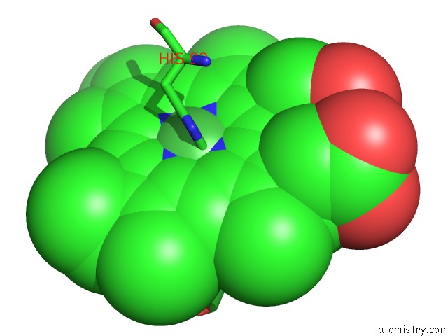
Mono view
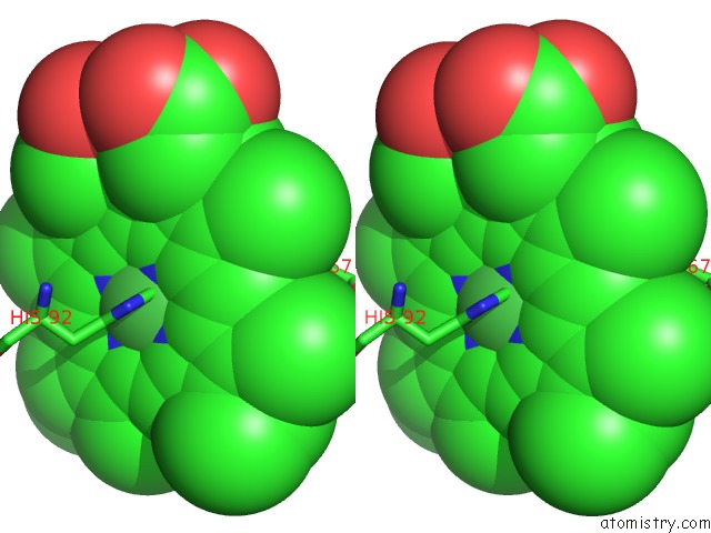
Stereo pair view
|
A full contact list of Nickel with other atoms in the Ni binding
site number 2 of Direct Observation of Conformational Population Shifts in Hemoglobin: Crystal Structure of Half-Liganded Hemoglobin After Adding 80 Mm Phosphate pH 6.7. within 5.0Å range:
probe
|
atom
|
residue
|
distance (Å)
|
B
|
Occ
|
D:Ni201
b:75.9
occ:1.00
|
NI
|
D:HNI201
|
0.0
|
75.9
|
1.0
|
NC
|
D:HNI201
|
2.0
|
75.0
|
1.0
|
NB
|
D:HNI201
|
2.0
|
74.5
|
1.0
|
NA
|
D:HNI201
|
2.0
|
77.0
|
1.0
|
ND
|
D:HNI201
|
2.0
|
77.6
|
1.0
|
NE2
|
D:HIS92
|
2.1
|
71.4
|
1.0
|
C1B
|
D:HNI201
|
3.0
|
75.0
|
1.0
|
C4B
|
D:HNI201
|
3.0
|
73.9
|
1.0
|
C1D
|
D:HNI201
|
3.0
|
78.2
|
1.0
|
C4C
|
D:HNI201
|
3.0
|
75.8
|
1.0
|
C1C
|
D:HNI201
|
3.0
|
74.0
|
1.0
|
CD2
|
D:HIS92
|
3.0
|
71.4
|
1.0
|
C4D
|
D:HNI201
|
3.0
|
79.2
|
1.0
|
C4A
|
D:HNI201
|
3.0
|
76.8
|
1.0
|
C1A
|
D:HNI201
|
3.1
|
78.5
|
1.0
|
CE1
|
D:HIS92
|
3.1
|
71.2
|
1.0
|
CHB
|
D:HNI201
|
3.4
|
76.3
|
1.0
|
CHC
|
D:HNI201
|
3.4
|
74.2
|
1.0
|
CHD
|
D:HNI201
|
3.4
|
77.8
|
1.0
|
CHA
|
D:HNI201
|
3.4
|
79.8
|
1.0
|
CG
|
D:HIS92
|
4.2
|
70.0
|
1.0
|
ND1
|
D:HIS92
|
4.2
|
71.9
|
1.0
|
C2B
|
D:HNI201
|
4.2
|
74.5
|
1.0
|
C3C
|
D:HNI201
|
4.2
|
75.5
|
1.0
|
C2C
|
D:HNI201
|
4.2
|
74.5
|
1.0
|
C3A
|
D:HNI201
|
4.3
|
78.3
|
1.0
|
C3B
|
D:HNI201
|
4.3
|
74.1
|
1.0
|
C2A
|
D:HNI201
|
4.3
|
79.3
|
1.0
|
C2D
|
D:HNI201
|
4.3
|
80.2
|
1.0
|
C3D
|
D:HNI201
|
4.3
|
80.9
|
1.0
|
NE2
|
D:HIS63
|
4.5
|
84.2
|
1.0
|
CG2
|
D:VAL67
|
4.8
|
76.2
|
1.0
|
|
Nickel binding site 3 out
of 6 in 5x2u
Go back to
Nickel Binding Sites List in 5x2u
Nickel binding site 3 out
of 6 in the Direct Observation of Conformational Population Shifts in Hemoglobin: Crystal Structure of Half-Liganded Hemoglobin After Adding 80 Mm Phosphate pH 6.7.
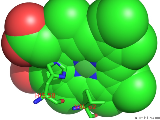
Mono view
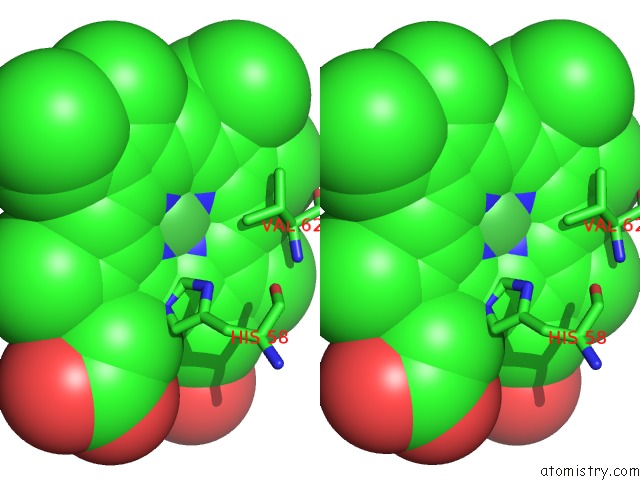
Stereo pair view
|
A full contact list of Nickel with other atoms in the Ni binding
site number 3 of Direct Observation of Conformational Population Shifts in Hemoglobin: Crystal Structure of Half-Liganded Hemoglobin After Adding 80 Mm Phosphate pH 6.7. within 5.0Å range:
probe
|
atom
|
residue
|
distance (Å)
|
B
|
Occ
|
E:Ni201
b:61.7
occ:1.00
|
NI
|
E:HNI201
|
0.0
|
61.7
|
1.0
|
ND
|
E:HNI201
|
1.9
|
62.9
|
1.0
|
NC
|
E:HNI201
|
2.0
|
61.4
|
1.0
|
NB
|
E:HNI201
|
2.0
|
60.6
|
1.0
|
NA
|
E:HNI201
|
2.0
|
62.2
|
1.0
|
NE2
|
E:HIS87
|
2.4
|
91.3
|
1.0
|
C1D
|
E:HNI201
|
2.9
|
63.7
|
1.0
|
C4B
|
E:HNI201
|
2.9
|
60.4
|
1.0
|
C4D
|
E:HNI201
|
3.0
|
63.9
|
1.0
|
C1C
|
E:HNI201
|
3.0
|
60.6
|
1.0
|
C4C
|
E:HNI201
|
3.0
|
62.1
|
1.0
|
C1B
|
E:HNI201
|
3.0
|
60.9
|
1.0
|
C4A
|
E:HNI201
|
3.0
|
61.9
|
1.0
|
C1A
|
E:HNI201
|
3.0
|
63.0
|
1.0
|
CHC
|
E:HNI201
|
3.3
|
60.8
|
1.0
|
CHD
|
E:HNI201
|
3.3
|
63.7
|
1.0
|
CE1
|
E:HIS87
|
3.4
|
91.1
|
1.0
|
CD2
|
E:HIS87
|
3.4
|
92.0
|
1.0
|
CHB
|
E:HNI201
|
3.4
|
61.7
|
1.0
|
CHA
|
E:HNI201
|
3.4
|
64.1
|
1.0
|
CE1
|
E:HIS58
|
4.1
|
83.6
|
1.0
|
C2D
|
E:HNI201
|
4.2
|
65.1
|
1.0
|
C3B
|
E:HNI201
|
4.2
|
60.5
|
1.0
|
C3C
|
E:HNI201
|
4.2
|
62.0
|
1.0
|
C2C
|
E:HNI201
|
4.2
|
61.2
|
1.0
|
C2B
|
E:HNI201
|
4.2
|
60.6
|
1.0
|
C3D
|
E:HNI201
|
4.3
|
65.3
|
1.0
|
C3A
|
E:HNI201
|
4.3
|
62.8
|
1.0
|
C2A
|
E:HNI201
|
4.3
|
63.5
|
1.0
|
ND1
|
E:HIS87
|
4.5
|
92.5
|
1.0
|
CG
|
E:HIS87
|
4.5
|
91.1
|
1.0
|
CG2
|
E:VAL62
|
4.7
|
66.8
|
1.0
|
NE2
|
E:HIS58
|
4.8
|
84.9
|
1.0
|
|
Nickel binding site 4 out
of 6 in 5x2u
Go back to
Nickel Binding Sites List in 5x2u
Nickel binding site 4 out
of 6 in the Direct Observation of Conformational Population Shifts in Hemoglobin: Crystal Structure of Half-Liganded Hemoglobin After Adding 80 Mm Phosphate pH 6.7.
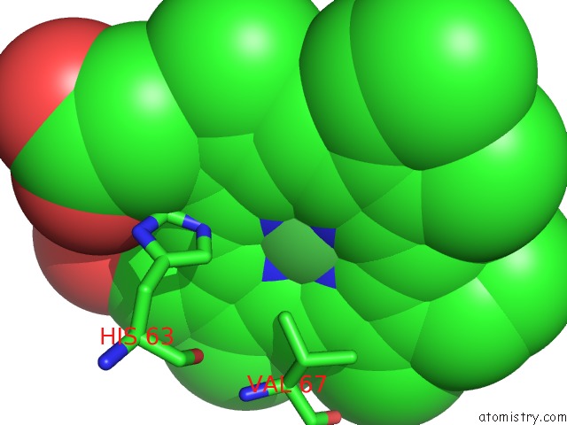
Mono view
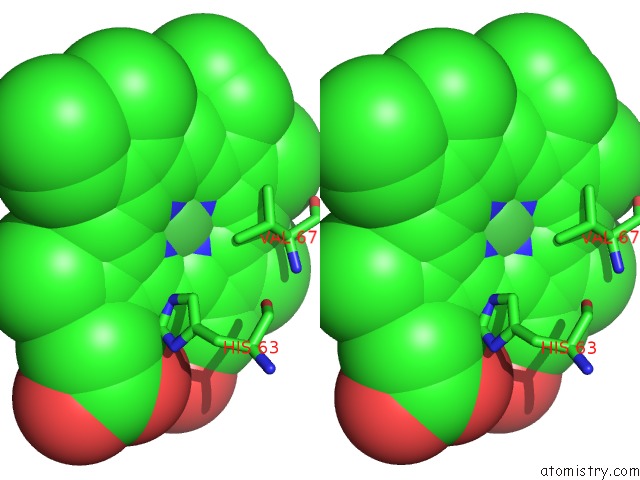
Stereo pair view
|
A full contact list of Nickel with other atoms in the Ni binding
site number 4 of Direct Observation of Conformational Population Shifts in Hemoglobin: Crystal Structure of Half-Liganded Hemoglobin After Adding 80 Mm Phosphate pH 6.7. within 5.0Å range:
probe
|
atom
|
residue
|
distance (Å)
|
B
|
Occ
|
H:Ni201
b:69.8
occ:1.00
|
NI
|
H:HNI201
|
0.0
|
69.8
|
1.0
|
NA
|
H:HNI201
|
1.9
|
70.3
|
1.0
|
NB
|
H:HNI201
|
2.0
|
68.5
|
1.0
|
ND
|
H:HNI201
|
2.0
|
71.1
|
1.0
|
NC
|
H:HNI201
|
2.0
|
69.5
|
1.0
|
NE2
|
H:HIS92
|
2.2
|
71.3
|
1.0
|
C4D
|
H:HNI201
|
3.0
|
72.2
|
1.0
|
C1B
|
H:HNI201
|
3.0
|
68.8
|
1.0
|
C1A
|
H:HNI201
|
3.0
|
71.3
|
1.0
|
C1D
|
H:HNI201
|
3.0
|
72.0
|
1.0
|
C4A
|
H:HNI201
|
3.0
|
70.0
|
1.0
|
C4B
|
H:HNI201
|
3.0
|
68.2
|
1.0
|
C4C
|
H:HNI201
|
3.0
|
70.4
|
1.0
|
C1C
|
H:HNI201
|
3.0
|
68.7
|
1.0
|
CE1
|
H:HIS92
|
3.1
|
71.3
|
1.0
|
CD2
|
H:HIS92
|
3.2
|
72.1
|
1.0
|
CHA
|
H:HNI201
|
3.4
|
72.4
|
1.0
|
CHB
|
H:HNI201
|
3.4
|
69.8
|
1.0
|
CHC
|
H:HNI201
|
3.4
|
68.7
|
1.0
|
CHD
|
H:HNI201
|
3.4
|
72.1
|
1.0
|
C2A
|
H:HNI201
|
4.2
|
71.8
|
1.0
|
C3A
|
H:HNI201
|
4.2
|
71.2
|
1.0
|
C2B
|
H:HNI201
|
4.2
|
68.4
|
1.0
|
C2C
|
H:HNI201
|
4.2
|
69.4
|
1.0
|
C3C
|
H:HNI201
|
4.3
|
70.4
|
1.0
|
C3D
|
H:HNI201
|
4.3
|
73.7
|
1.0
|
C2D
|
H:HNI201
|
4.3
|
73.6
|
1.0
|
ND1
|
H:HIS92
|
4.3
|
72.8
|
1.0
|
C3B
|
H:HNI201
|
4.3
|
68.3
|
1.0
|
CG
|
H:HIS92
|
4.3
|
71.1
|
1.0
|
CG2
|
H:VAL67
|
4.3
|
77.8
|
1.0
|
NE2
|
H:HIS63
|
4.6
|
81.1
|
1.0
|
|
Nickel binding site 5 out
of 6 in 5x2u
Go back to
Nickel Binding Sites List in 5x2u
Nickel binding site 5 out
of 6 in the Direct Observation of Conformational Population Shifts in Hemoglobin: Crystal Structure of Half-Liganded Hemoglobin After Adding 80 Mm Phosphate pH 6.7.
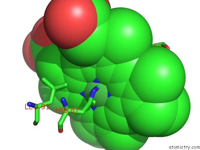
Mono view
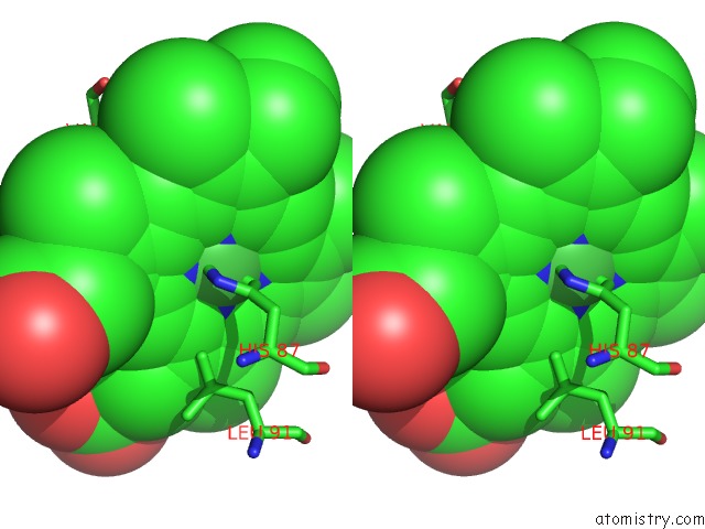
Stereo pair view
|
A full contact list of Nickel with other atoms in the Ni binding
site number 5 of Direct Observation of Conformational Population Shifts in Hemoglobin: Crystal Structure of Half-Liganded Hemoglobin After Adding 80 Mm Phosphate pH 6.7. within 5.0Å range:
probe
|
atom
|
residue
|
distance (Å)
|
B
|
Occ
|
I:Ni201
b:60.0
occ:1.00
|
NI
|
I:HNI201
|
0.0
|
60.0
|
1.0
|
NB
|
I:HNI201
|
2.0
|
59.1
|
1.0
|
ND
|
I:HNI201
|
2.0
|
61.2
|
1.0
|
NC
|
I:HNI201
|
2.0
|
59.5
|
1.0
|
NA
|
I:HNI201
|
2.0
|
60.9
|
1.0
|
NE2
|
I:HIS87
|
2.3
|
87.2
|
1.0
|
CE1
|
I:HIS87
|
2.9
|
87.0
|
1.0
|
C4B
|
I:HNI201
|
3.0
|
58.9
|
1.0
|
C1D
|
I:HNI201
|
3.0
|
61.7
|
1.0
|
C4D
|
I:HNI201
|
3.0
|
62.3
|
1.0
|
C1B
|
I:HNI201
|
3.0
|
59.5
|
1.0
|
C1C
|
I:HNI201
|
3.0
|
58.8
|
1.0
|
C4C
|
I:HNI201
|
3.0
|
60.0
|
1.0
|
C1A
|
I:HNI201
|
3.0
|
61.8
|
1.0
|
C4A
|
I:HNI201
|
3.0
|
60.6
|
1.0
|
CHC
|
I:HNI201
|
3.3
|
59.3
|
1.0
|
CHD
|
I:HNI201
|
3.4
|
61.5
|
1.0
|
CHA
|
I:HNI201
|
3.4
|
62.9
|
1.0
|
CHB
|
I:HNI201
|
3.4
|
60.4
|
1.0
|
CD2
|
I:HIS87
|
3.6
|
87.7
|
1.0
|
CE1
|
I:HIS58
|
4.0
|
71.4
|
1.0
|
NE2
|
I:HIS58
|
4.1
|
72.2
|
1.0
|
ND1
|
I:HIS87
|
4.2
|
88.2
|
1.0
|
C2D
|
I:HNI201
|
4.2
|
63.1
|
1.0
|
C3B
|
I:HNI201
|
4.2
|
59.2
|
1.0
|
C2A
|
I:HNI201
|
4.2
|
62.5
|
1.0
|
C3D
|
I:HNI201
|
4.2
|
63.6
|
1.0
|
C3A
|
I:HNI201
|
4.2
|
61.8
|
1.0
|
C2B
|
I:HNI201
|
4.2
|
59.2
|
1.0
|
C3C
|
I:HNI201
|
4.2
|
59.8
|
1.0
|
C2C
|
I:HNI201
|
4.3
|
59.2
|
1.0
|
CG
|
I:HIS87
|
4.5
|
86.5
|
1.0
|
CG2
|
I:VAL62
|
4.6
|
69.8
|
1.0
|
CD2
|
I:LEU91
|
4.9
|
77.9
|
1.0
|
|
Nickel binding site 6 out
of 6 in 5x2u
Go back to
Nickel Binding Sites List in 5x2u
Nickel binding site 6 out
of 6 in the Direct Observation of Conformational Population Shifts in Hemoglobin: Crystal Structure of Half-Liganded Hemoglobin After Adding 80 Mm Phosphate pH 6.7.
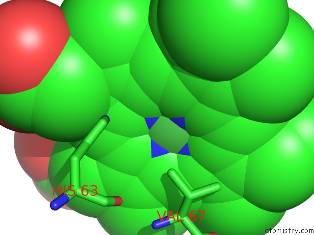
Mono view
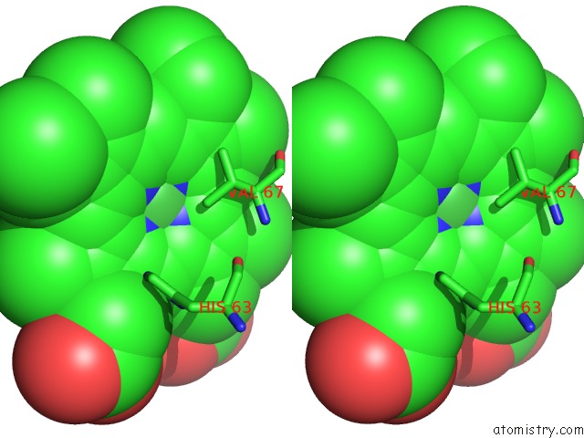
Stereo pair view
|
A full contact list of Nickel with other atoms in the Ni binding
site number 6 of Direct Observation of Conformational Population Shifts in Hemoglobin: Crystal Structure of Half-Liganded Hemoglobin After Adding 80 Mm Phosphate pH 6.7. within 5.0Å range:
probe
|
atom
|
residue
|
distance (Å)
|
B
|
Occ
|
L:Ni201
b:74.2
occ:1.00
|
NI
|
L:HNI201
|
0.0
|
74.2
|
1.0
|
NA
|
L:HNI201
|
2.0
|
75.0
|
1.0
|
NC
|
L:HNI201
|
2.0
|
73.6
|
1.0
|
ND
|
L:HNI201
|
2.0
|
75.5
|
1.0
|
NB
|
L:HNI201
|
2.0
|
72.9
|
1.0
|
NE2
|
L:HIS92
|
2.1
|
71.0
|
1.0
|
CE1
|
L:HIS92
|
3.0
|
70.7
|
1.0
|
C1D
|
L:HNI201
|
3.0
|
76.2
|
1.0
|
C1B
|
L:HNI201
|
3.0
|
73.2
|
1.0
|
C4C
|
L:HNI201
|
3.0
|
74.4
|
1.0
|
C1C
|
L:HNI201
|
3.0
|
72.7
|
1.0
|
C4B
|
L:HNI201
|
3.0
|
72.5
|
1.0
|
C4A
|
L:HNI201
|
3.0
|
74.6
|
1.0
|
C4D
|
L:HNI201
|
3.0
|
76.8
|
1.0
|
C1A
|
L:HNI201
|
3.0
|
76.0
|
1.0
|
CD2
|
L:HIS92
|
3.1
|
72.0
|
1.0
|
CHB
|
L:HNI201
|
3.4
|
74.3
|
1.0
|
CHD
|
L:HNI201
|
3.4
|
76.1
|
1.0
|
CHC
|
L:HNI201
|
3.4
|
72.8
|
1.0
|
CHA
|
L:HNI201
|
3.4
|
77.2
|
1.0
|
ND1
|
L:HIS92
|
4.1
|
72.2
|
1.0
|
CG
|
L:HIS92
|
4.2
|
71.1
|
1.0
|
C2C
|
L:HNI201
|
4.2
|
73.4
|
1.0
|
C2A
|
L:HNI201
|
4.2
|
76.7
|
1.0
|
C3A
|
L:HNI201
|
4.2
|
75.9
|
1.0
|
C2B
|
L:HNI201
|
4.3
|
72.8
|
1.0
|
C3C
|
L:HNI201
|
4.3
|
74.2
|
1.0
|
C2D
|
L:HNI201
|
4.3
|
77.9
|
1.0
|
C3B
|
L:HNI201
|
4.3
|
72.6
|
1.0
|
C3D
|
L:HNI201
|
4.3
|
78.3
|
1.0
|
CG2
|
L:VAL67
|
4.5
|
83.0
|
1.0
|
NE2
|
L:HIS63
|
4.9
|
92.8
|
1.0
|
|
Reference:
N.Shibayama,
M.Ohki,
J.R.H.Tame,
S.Y.Park.
Direct Observation of Conformational Population Shifts in Crystalline Human Hemoglobin. J. Biol. Chem. V. 292 18258 2017.
ISSN: ESSN 1083-351X
PubMed: 28931607
DOI: 10.1074/JBC.M117.781146
Page generated: Thu Oct 10 08:10:42 2024
|