Nickel »
PDB 6vwz-6z8m »
6x5e »
Nickel in PDB 6x5e: Crystal Structure of A Lewis-Binding Fab (CH88.2)
Protein crystallography data
The structure of Crystal Structure of A Lewis-Binding Fab (CH88.2), PDB code: 6x5e
was solved by
C.Soliman,
P.A.Ramsland,
with X-Ray Crystallography technique. A brief refinement statistics is given in the table below:
| Resolution Low / High (Å) | 33.49 / 2.29 |
| Space group | P 1 |
| Cell size a, b, c (Å), α, β, γ (°) | 38.423, 68.480, 91.750, 110.50, 99.29, 90.01 |
| R / Rfree (%) | 23 / 28.4 |
Nickel Binding Sites:
The binding sites of Nickel atom in the Crystal Structure of A Lewis-Binding Fab (CH88.2)
(pdb code 6x5e). This binding sites where shown within
5.0 Angstroms radius around Nickel atom.
In total 3 binding sites of Nickel where determined in the Crystal Structure of A Lewis-Binding Fab (CH88.2), PDB code: 6x5e:
Jump to Nickel binding site number: 1; 2; 3;
In total 3 binding sites of Nickel where determined in the Crystal Structure of A Lewis-Binding Fab (CH88.2), PDB code: 6x5e:
Jump to Nickel binding site number: 1; 2; 3;
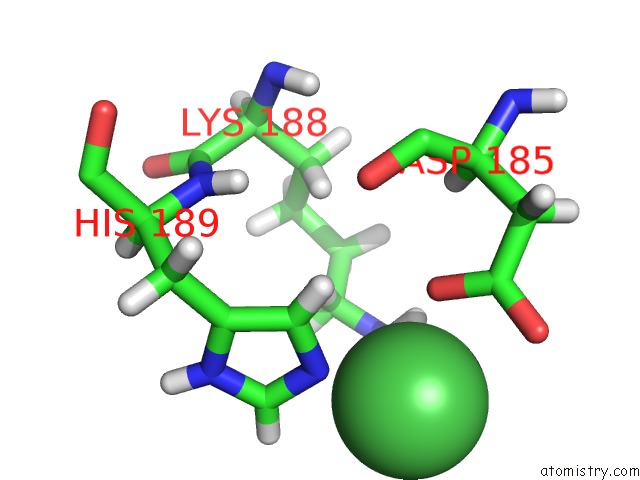
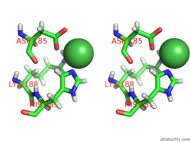
Nickel binding site 1 out of 3 in 6x5e
Go back to
Nickel binding site 1 out
of 3 in the Crystal Structure of A Lewis-Binding Fab (CH88.2)
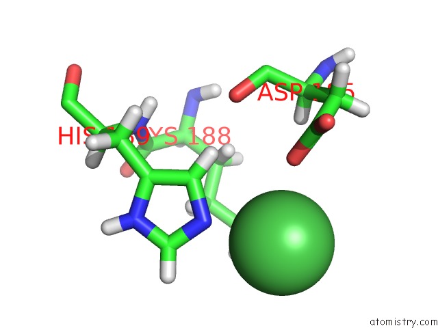
Mono view
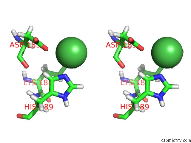
Stereo pair view
Mono view
Stereo pair view
A full contact list of Nickel with other atoms in the Ni binding
site number 1 of Crystal Structure of A Lewis-Binding Fab (CH88.2) within 5.0Å range:
|
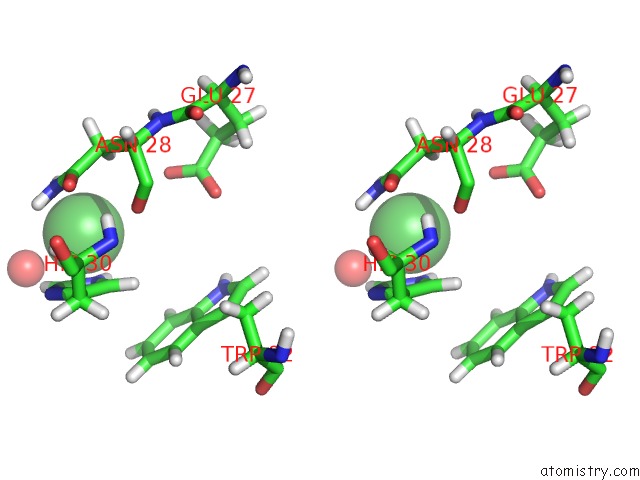
Nickel binding site 2 out of 3 in 6x5e
Go back to
Nickel binding site 2 out
of 3 in the Crystal Structure of A Lewis-Binding Fab (CH88.2)
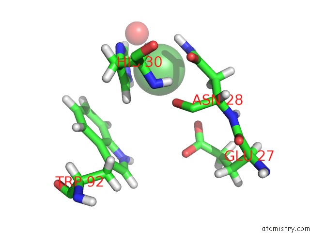
Mono view
Stereo pair view
Mono view
Stereo pair view
A full contact list of Nickel with other atoms in the Ni binding
site number 2 of Crystal Structure of A Lewis-Binding Fab (CH88.2) within 5.0Å range:
|
Nickel binding site 3 out of 3 in 6x5e
Go back to
Nickel binding site 3 out
of 3 in the Crystal Structure of A Lewis-Binding Fab (CH88.2)
Mono view
Stereo pair view
Mono view
Stereo pair view
A full contact list of Nickel with other atoms in the Ni binding
site number 3 of Crystal Structure of A Lewis-Binding Fab (CH88.2) within 5.0Å range:
|
Reference:
C.Soliman,
A.J.Guy,
J.X.Chua,
M.Vankemmelbeke,
R.S.Mcintosh,
S.Eastwood,
V.K.Truong,
A.Elbourne,
I.Spendlove,
L.G.Durrant,
P.A.Ramsland.
Molecular and Structural Basis For Lewis Glycan Recognition By A Cancer-Targeting Antibody. Biochem.J. V. 477 3219 2020.
ISSN: ESSN 1470-8728
PubMed: 32789497
DOI: 10.1042/BCJ20200454
Page generated: Thu Oct 10 08:58:25 2024
ISSN: ESSN 1470-8728
PubMed: 32789497
DOI: 10.1042/BCJ20200454
Last articles
Zn in 9MJ5Zn in 9HNW
Zn in 9G0L
Zn in 9FNE
Zn in 9DZN
Zn in 9E0I
Zn in 9D32
Zn in 9DAK
Zn in 8ZXC
Zn in 8ZUF