Nickel »
PDB 6vwz-6z8m »
6yag »
Nickel in PDB 6yag: Crystal Structure of Pnra From S. Pneumoniae in Complex with Thymidine
Protein crystallography data
The structure of Crystal Structure of Pnra From S. Pneumoniae in Complex with Thymidine, PDB code: 6yag
was solved by
M.T.Batuecas,
J.A.Hermoso,
with X-Ray Crystallography technique. A brief refinement statistics is given in the table below:
| Resolution Low / High (Å) | 49.21 / 2.55 |
| Space group | C 2 2 21 |
| Cell size a, b, c (Å), α, β, γ (°) | 78.966, 303.818, 128.417, 90, 90, 90 |
| R / Rfree (%) | 20.9 / 25.5 |
Nickel Binding Sites:
The binding sites of Nickel atom in the Crystal Structure of Pnra From S. Pneumoniae in Complex with Thymidine
(pdb code 6yag). This binding sites where shown within
5.0 Angstroms radius around Nickel atom.
In total 7 binding sites of Nickel where determined in the Crystal Structure of Pnra From S. Pneumoniae in Complex with Thymidine, PDB code: 6yag:
Jump to Nickel binding site number: 1; 2; 3; 4; 5; 6; 7;
In total 7 binding sites of Nickel where determined in the Crystal Structure of Pnra From S. Pneumoniae in Complex with Thymidine, PDB code: 6yag:
Jump to Nickel binding site number: 1; 2; 3; 4; 5; 6; 7;
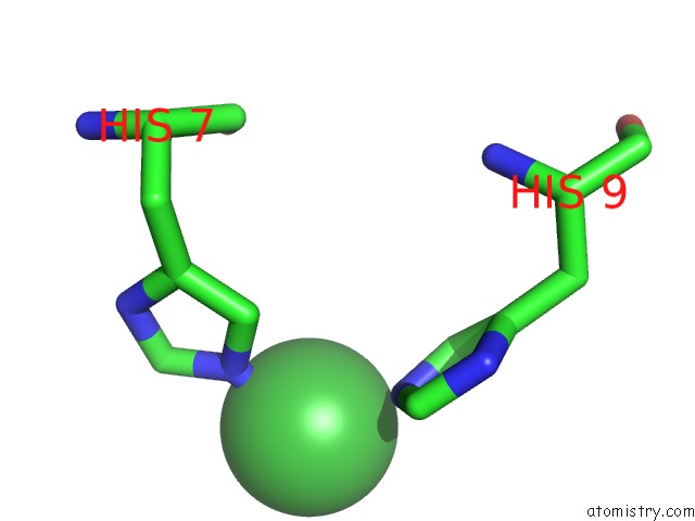
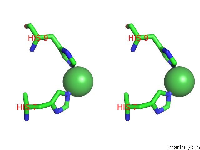
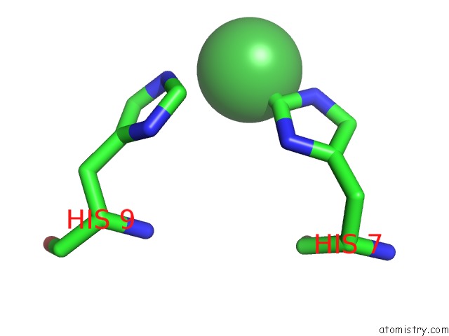
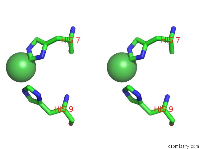
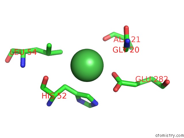
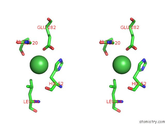
Nickel binding site 1 out of 7 in 6yag
Go back to
Nickel binding site 1 out
of 7 in the Crystal Structure of Pnra From S. Pneumoniae in Complex with Thymidine
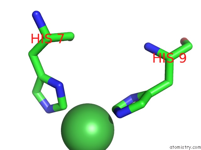
Mono view
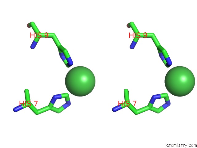
Stereo pair view
Mono view
Stereo pair view
A full contact list of Nickel with other atoms in the Ni binding
site number 1 of Crystal Structure of Pnra From S. Pneumoniae in Complex with Thymidine within 5.0Å range:
|
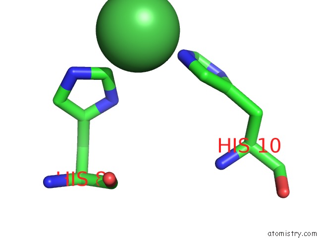
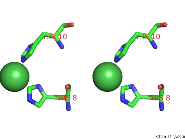
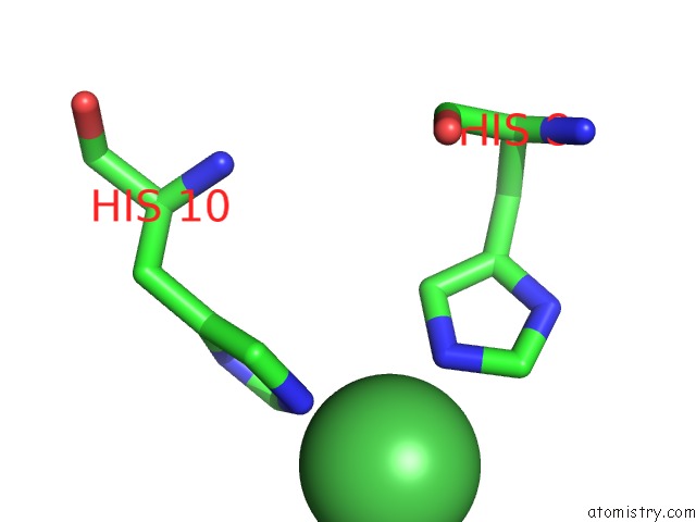
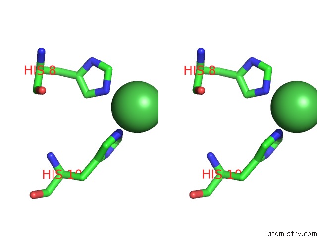
Nickel binding site 2 out of 7 in 6yag
Go back to
Nickel binding site 2 out
of 7 in the Crystal Structure of Pnra From S. Pneumoniae in Complex with Thymidine
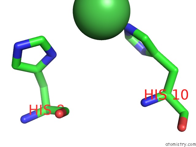
Mono view
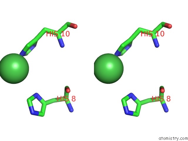
Stereo pair view
Mono view
Stereo pair view
A full contact list of Nickel with other atoms in the Ni binding
site number 2 of Crystal Structure of Pnra From S. Pneumoniae in Complex with Thymidine within 5.0Å range:
|
Nickel binding site 3 out of 7 in 6yag
Go back to
Nickel binding site 3 out
of 7 in the Crystal Structure of Pnra From S. Pneumoniae in Complex with Thymidine
Mono view
Stereo pair view
Mono view
Stereo pair view
A full contact list of Nickel with other atoms in the Ni binding
site number 3 of Crystal Structure of Pnra From S. Pneumoniae in Complex with Thymidine within 5.0Å range:
|
Nickel binding site 4 out of 7 in 6yag
Go back to
Nickel binding site 4 out
of 7 in the Crystal Structure of Pnra From S. Pneumoniae in Complex with Thymidine
Mono view
Stereo pair view
Mono view
Stereo pair view
A full contact list of Nickel with other atoms in the Ni binding
site number 4 of Crystal Structure of Pnra From S. Pneumoniae in Complex with Thymidine within 5.0Å range:
|
Nickel binding site 5 out of 7 in 6yag
Go back to
Nickel binding site 5 out
of 7 in the Crystal Structure of Pnra From S. Pneumoniae in Complex with Thymidine
Mono view
Stereo pair view
Mono view
Stereo pair view
A full contact list of Nickel with other atoms in the Ni binding
site number 5 of Crystal Structure of Pnra From S. Pneumoniae in Complex with Thymidine within 5.0Å range:
|
Nickel binding site 6 out of 7 in 6yag
Go back to
Nickel binding site 6 out
of 7 in the Crystal Structure of Pnra From S. Pneumoniae in Complex with Thymidine
Mono view
Stereo pair view
Mono view
Stereo pair view
A full contact list of Nickel with other atoms in the Ni binding
site number 6 of Crystal Structure of Pnra From S. Pneumoniae in Complex with Thymidine within 5.0Å range:
|
Nickel binding site 7 out of 7 in 6yag
Go back to
Nickel binding site 7 out
of 7 in the Crystal Structure of Pnra From S. Pneumoniae in Complex with Thymidine
Mono view
Stereo pair view
Mono view
Stereo pair view
A full contact list of Nickel with other atoms in the Ni binding
site number 7 of Crystal Structure of Pnra From S. Pneumoniae in Complex with Thymidine within 5.0Å range:
|
Reference:
M.R.Abdullah,
M.T.Batuecas,
F.Jennert,
F.Voss,
P.Westhoff,
T.P.Kohler,
R.Molina,
S.Hirschmann,
M.Lalk,
J.A.Hermoso,
S.Hammerschmidt.
Crystal Structure and Pathophysiological Role of the Pneumococcal Nucleoside-Binding Protein Pnra. J.Mol.Biol. V. 433 66723 2021.
ISSN: ESSN 1089-8638
PubMed: 33242497
DOI: 10.1016/J.JMB.2020.11.022
Page generated: Thu Oct 10 09:00:27 2024
ISSN: ESSN 1089-8638
PubMed: 33242497
DOI: 10.1016/J.JMB.2020.11.022
Last articles
Zn in 9J0NZn in 9J0O
Zn in 9J0P
Zn in 9FJX
Zn in 9EKB
Zn in 9C0F
Zn in 9CAH
Zn in 9CH0
Zn in 9CH3
Zn in 9CH1