Nickel »
PDB 6z8o-7err »
7b1s »
Nickel in PDB 7b1s: Crystal Structure of the Ethyl-Coenzyme M Reductase From Candidatus Ethanoperedens Thermophilum at 0.994-A Resolution
Enzymatic activity of Crystal Structure of the Ethyl-Coenzyme M Reductase From Candidatus Ethanoperedens Thermophilum at 0.994-A Resolution
All present enzymatic activity of Crystal Structure of the Ethyl-Coenzyme M Reductase From Candidatus Ethanoperedens Thermophilum at 0.994-A Resolution:
2.8.4.1;
2.8.4.1;
Protein crystallography data
The structure of Crystal Structure of the Ethyl-Coenzyme M Reductase From Candidatus Ethanoperedens Thermophilum at 0.994-A Resolution, PDB code: 7b1s
was solved by
T.Wagner,
O.N.Lemaire,
S.Engilberge,
with X-Ray Crystallography technique. A brief refinement statistics is given in the table below:
| Resolution Low / High (Å) | 39.76 / 0.99 |
| Space group | P 1 21 1 |
| Cell size a, b, c (Å), α, β, γ (°) | 83.736, 146.927, 113.128, 90, 106.98, 90 |
| R / Rfree (%) | 11.2 / 12.8 |
Other elements in 7b1s:
The structure of Crystal Structure of the Ethyl-Coenzyme M Reductase From Candidatus Ethanoperedens Thermophilum at 0.994-A Resolution also contains other interesting chemical elements:
| Potassium | (K) | 4 atoms |
| Chlorine | (Cl) | 6 atoms |
| Manganese | (Mn) | 1 atom |
Nickel Binding Sites:
The binding sites of Nickel atom in the Crystal Structure of the Ethyl-Coenzyme M Reductase From Candidatus Ethanoperedens Thermophilum at 0.994-A Resolution
(pdb code 7b1s). This binding sites where shown within
5.0 Angstroms radius around Nickel atom.
In total 2 binding sites of Nickel where determined in the Crystal Structure of the Ethyl-Coenzyme M Reductase From Candidatus Ethanoperedens Thermophilum at 0.994-A Resolution, PDB code: 7b1s:
Jump to Nickel binding site number: 1; 2;
In total 2 binding sites of Nickel where determined in the Crystal Structure of the Ethyl-Coenzyme M Reductase From Candidatus Ethanoperedens Thermophilum at 0.994-A Resolution, PDB code: 7b1s:
Jump to Nickel binding site number: 1; 2;
Nickel binding site 1 out of 2 in 7b1s
Go back to
Nickel binding site 1 out
of 2 in the Crystal Structure of the Ethyl-Coenzyme M Reductase From Candidatus Ethanoperedens Thermophilum at 0.994-A Resolution
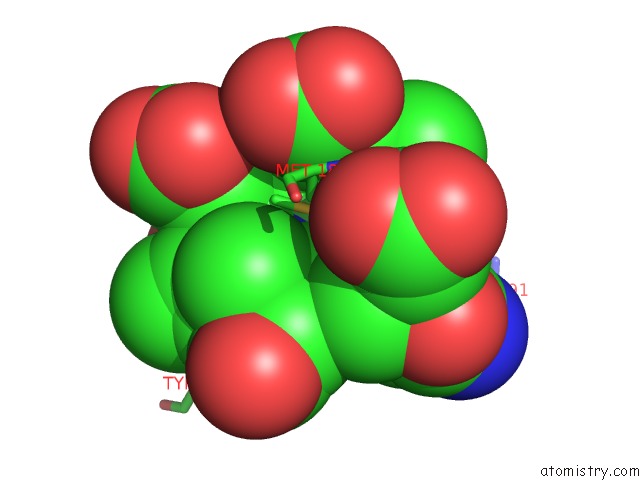
Mono view
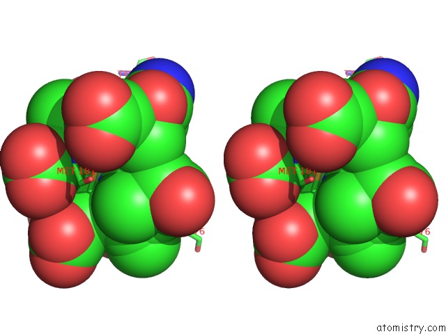
Stereo pair view
Mono view
Stereo pair view
A full contact list of Nickel with other atoms in the Ni binding
site number 1 of Crystal Structure of the Ethyl-Coenzyme M Reductase From Candidatus Ethanoperedens Thermophilum at 0.994-A Resolution within 5.0Å range:
|
Nickel binding site 2 out of 2 in 7b1s
Go back to
Nickel binding site 2 out
of 2 in the Crystal Structure of the Ethyl-Coenzyme M Reductase From Candidatus Ethanoperedens Thermophilum at 0.994-A Resolution
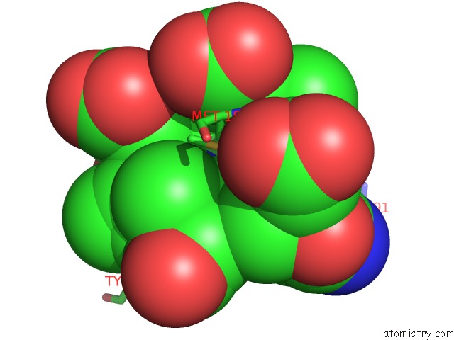
Mono view
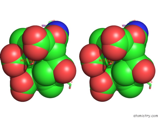
Stereo pair view
Mono view
Stereo pair view
A full contact list of Nickel with other atoms in the Ni binding
site number 2 of Crystal Structure of the Ethyl-Coenzyme M Reductase From Candidatus Ethanoperedens Thermophilum at 0.994-A Resolution within 5.0Å range:
|
Reference:
C.J.Hahn,
O.N.Lemaire,
J.Kahnt,
S.Engilberge,
G.Wegener,
T.Wagner.
Crystal Structure of A Key Enzyme For Anaerobic Ethane Activation. Science V. 373 118 2021.
ISSN: ESSN 1095-9203
PubMed: 34210888
DOI: 10.1126/SCIENCE.ABG1765
Page generated: Thu Oct 10 09:06:32 2024
ISSN: ESSN 1095-9203
PubMed: 34210888
DOI: 10.1126/SCIENCE.ABG1765
Last articles
Zn in 9MJ5Zn in 9HNW
Zn in 9G0L
Zn in 9FNE
Zn in 9DZN
Zn in 9E0I
Zn in 9D32
Zn in 9DAK
Zn in 8ZXC
Zn in 8ZUF