Nickel »
PDB 7odh-7utd »
7rei »
Nickel in PDB 7rei: The Crystal Structure of Nickel Bound Human Ado C18S C239S Variant
Enzymatic activity of The Crystal Structure of Nickel Bound Human Ado C18S C239S Variant
All present enzymatic activity of The Crystal Structure of Nickel Bound Human Ado C18S C239S Variant:
1.13.11.19;
1.13.11.19;
Protein crystallography data
The structure of The Crystal Structure of Nickel Bound Human Ado C18S C239S Variant, PDB code: 7rei
was solved by
Y.Wang,
I.Shin,
J.Li,
A.Liu,
with X-Ray Crystallography technique. A brief refinement statistics is given in the table below:
| Resolution Low / High (Å) | 47.62 / 1.78 |
| Space group | C 2 2 21 |
| Cell size a, b, c (Å), α, β, γ (°) | 54.881, 95.764, 117.614, 90, 90, 90 |
| R / Rfree (%) | 18 / 20.9 |
Nickel Binding Sites:
The binding sites of Nickel atom in the The Crystal Structure of Nickel Bound Human Ado C18S C239S Variant
(pdb code 7rei). This binding sites where shown within
5.0 Angstroms radius around Nickel atom.
In total only one binding site of Nickel was determined in the The Crystal Structure of Nickel Bound Human Ado C18S C239S Variant, PDB code: 7rei:
In total only one binding site of Nickel was determined in the The Crystal Structure of Nickel Bound Human Ado C18S C239S Variant, PDB code: 7rei:
Nickel binding site 1 out of 1 in 7rei
Go back to
Nickel binding site 1 out
of 1 in the The Crystal Structure of Nickel Bound Human Ado C18S C239S Variant
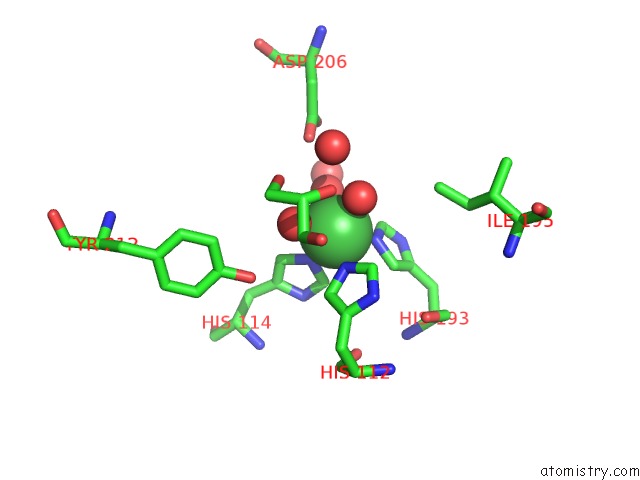
Mono view
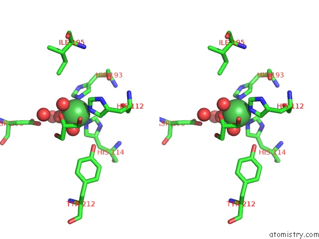
Stereo pair view
Mono view
Stereo pair view
A full contact list of Nickel with other atoms in the Ni binding
site number 1 of The Crystal Structure of Nickel Bound Human Ado C18S C239S Variant within 5.0Å range:
|
Reference:
Y.Wang,
I.Shin,
J.Li,
A.Liu.
Crystal Structure of Human Cysteamine Dioxygenase Provides A Structural Rationale For Its Function As An Oxygen Sensor J.Biol.Chem. 2021.
ISSN: ESSN 1083-351X
DOI: 10.1016/J.JBC.2021.101176
Page generated: Mon Aug 18 21:58:15 2025
ISSN: ESSN 1083-351X
DOI: 10.1016/J.JBC.2021.101176
Last articles
K in 9CWUK in 9CVB
K in 9CVA
K in 9COM
Fe in 9VR0
Fe in 9UD8
Fe in 9QDT
Fe in 9S2T
Fe in 9JQA
Fe in 9IYV