Nickel »
PDB 3qsi-3tsn »
3slu »
Nickel in PDB 3slu: Crystal Structure of NMB0315
Protein crystallography data
The structure of Crystal Structure of NMB0315, PDB code: 3slu
was solved by
Y.Shen,
X.Wang,
X.Yang,
H.Xu,
with X-Ray Crystallography technique. A brief refinement statistics is given in the table below:
| Resolution Low / High (Å) | 37.75 / 2.41 |
| Space group | C 1 2 1 |
| Cell size a, b, c (Å), α, β, γ (°) | 195.680, 75.500, 81.640, 90.00, 94.66, 90.00 |
| R / Rfree (%) | 25.5 / 26.4 |
Nickel Binding Sites:
The binding sites of Nickel atom in the Crystal Structure of NMB0315
(pdb code 3slu). This binding sites where shown within
5.0 Angstroms radius around Nickel atom.
In total 2 binding sites of Nickel where determined in the Crystal Structure of NMB0315, PDB code: 3slu:
Jump to Nickel binding site number: 1; 2;
In total 2 binding sites of Nickel where determined in the Crystal Structure of NMB0315, PDB code: 3slu:
Jump to Nickel binding site number: 1; 2;
Nickel binding site 1 out of 2 in 3slu
Go back to
Nickel binding site 1 out
of 2 in the Crystal Structure of NMB0315
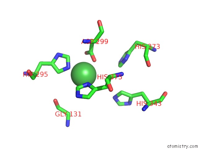
Mono view
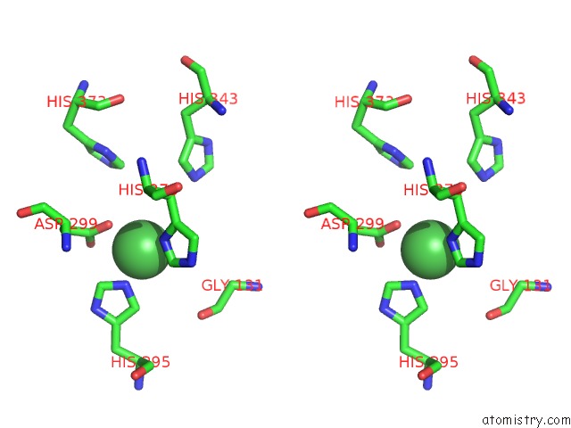
Stereo pair view
Mono view
Stereo pair view
A full contact list of Nickel with other atoms in the Ni binding
site number 1 of Crystal Structure of NMB0315 within 5.0Å range:
|
Nickel binding site 2 out of 2 in 3slu
Go back to
Nickel binding site 2 out
of 2 in the Crystal Structure of NMB0315
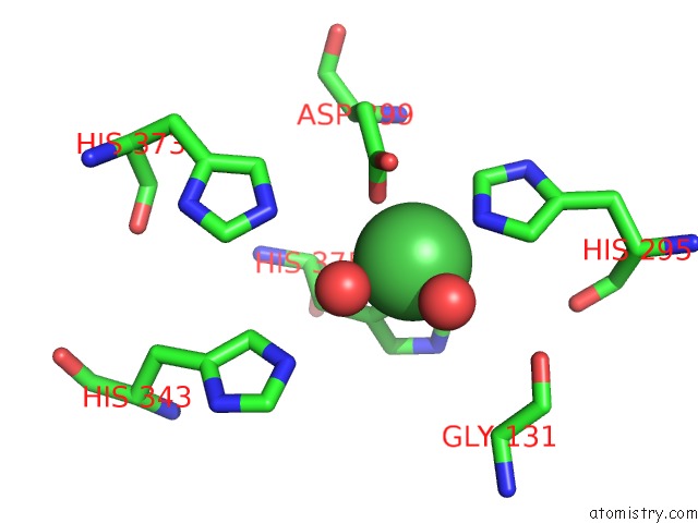
Mono view
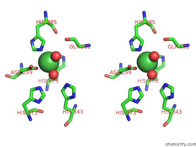
Stereo pair view
Mono view
Stereo pair view
A full contact list of Nickel with other atoms in the Ni binding
site number 2 of Crystal Structure of NMB0315 within 5.0Å range:
|
Reference:
X.Wang,
X.Yang,
C.Yang,
Z.Wu,
H.Xu,
Y.Shen.
Crystal Structure of Outer Membrane Protein NMB0315 From Neisseria Meningitidis. Plos One V. 6 26845 2011.
ISSN: ESSN 1932-6203
PubMed: 22046377
DOI: 10.1371/JOURNAL.PONE.0026845
Page generated: Mon Aug 18 18:58:41 2025
ISSN: ESSN 1932-6203
PubMed: 22046377
DOI: 10.1371/JOURNAL.PONE.0026845
Last articles
Ni in 5WK0Ni in 5WAY
Ni in 5WJA
Ni in 5VMP
Ni in 5VGI
Ni in 5VB0
Ni in 5VP3
Ni in 5UDW
Ni in 5VFB
Ni in 5V9T